
Communication network in the human brain
The brain is an extremely important part of the body, there are about 100 billion neurons in the brain involved in controlling the functions of organs and parts in the body. With such a large number of cells, how does the brain communicate and how are nerve signals transmitted?
1. How does the brain communicate?
Neurons in the brain communicate through a combination of electrical and chemical signals. In neurons, electrical signals controlled by charged particles allow rapid conduction from one end of the cell to the other.
Communication between neurons occurs at small gaps called synapses. There are specialized divisions of two cells (i.e. presynaptic and postsynaptic neurons) spaced just nanometers apart to allow for chemical transmission.
Presynaptic neurons release a chemical - a neurotransmitter. Postsynaptic neurons have specialized proteins called neurotransmitter receptors that receive these neurotransmitters. Neurotransmitter molecules bind to receptor proteins and alter postsynaptic neuron function.
Two types of neurotransmitter receptors coexist: ion channels, which allow the rapid flow of ions directly across the cell membrane to the outside, and receptors that combine with the G protein, setting the events send chemical signals to move in the cell.
There are hundreds of molecules known to act as neurotransmitters in the brain. The development and function of neurons are also affected by peptides called neurotrophins and by steroid hormones.
2. Factors involved in nerve signal transmission in the brain
2.1. Neuron-to-Neuro communication
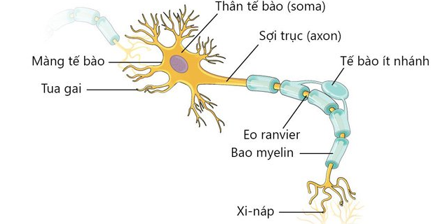
Neurons are cells in the brain that are responsible for transmitting information quickly. Nerve cells are specially constructed that give them properties that allow them to perform their unique role in the nervous system.
The shape of a neuron is one such unique feature. In addition to the cell body, or soma, like that of other cells, neurons have specialized thin branches called dendrites and axons. Neurons receive chemical input from other neurons via dendrites and transmit information to other cells via axons.
Neurons are also “excitable” cells. The surface of nerve cell membranes contains numerous proteins called ion channels that allow small electrically charged atoms to pass from one side of the membrane to the other. Some of these channels are opened when the voltage across the cell membrane changes. A subtype of these "potential" channels allow neurons to generate a rapid signal called an "action potential," which is the fastest form of intracellular electrical signal transmission in biology. .
Individual neurons are usually completely separated from each other by the outer cell membrane, so electrical or chemical signals cannot be shared directly. Signaling is carried out only at the electrical synapse, where ion pathways are made of so-called junction proteins that connect the intracellular compartments of neighboring neurons, allowing the flow of ions. directly from cell to cell. This form of nerve communication is less common in the central nervous system than chemical transmission.
Communication in the human brain is mainly chemical communication between neurons that involves the release of neurotransmitters from a neuron, altering the activity of the next neuron. receive. This chemical communication usually occurs at a specialized structure called a synapse, where parts of two cells are spaced between 20 and 50 nanometers apart.
Neurons that release chemicals are called presynaptic neurons. A special structure at the axon terminal of a presynaptic neuron, called the axon terminal, contains small packages called vesicles, which are filled with neurotransmitter molecules. When an action potential reaches the axon terminal and stimulates an increase in calcium concentration, this ion stimulates the vesicle to fuse with the cell membrane and release the neurotransmitter into the small synaptic space between the cells. cell.
Neurons that are acted on by chemicals are called postsynaptic neurons. Neurotransmitter molecules released from presynaptic vesicles cross the synaptic cleft and bind to proteins, called neurotransmitter receptors, on the surface membranes of postsynaptic neurons.
Neurotransmitters are rapidly cleared from the synapse once they are released. This minimizes the time they take to interact with the receptors so that discrete, short synaptic signals are generated. At most synapses in the brain, specific proteins called neurotransmitters mediate this removal.
Transport proteins reside in cell surface membranes and actively move neurotransmitter molecules from the outside to the inside of the cell. In many cases, this uptake occurs at the very terminal of the synapse, where the neurotransmitter is loaded directly into the vesicles.
However, in some cases, helper cells i.e. glial cells are also involved in neurotransmitter uptake. Elimination of synaptic neurotransmitters can also occur through enzymes that degrade the neurotransmitter into constitutive molecules that do not activate receptors on their own.

2.2. Neurotransmitter receptor
Neurotransmitter receptors are divided into two main categories, including:
Ligand ion channel receptors (LGICs): are specialized proteins that rapidly transmit chemical signals that transmit neurotransmitters directly into responses electricity. Part of the protein is specialized for binding to the neurotransmitter molecule. This "binding site" is located in the protein outside the cell. The part of the protein that is buried in the cell surface membrane forms an ion hole, which is essentially a fluid-filled tunnel across the membrane through which charged ions can pass. The time from neurotransmitter binding to ion hole opening ranges from microseconds to milliseconds.
Thus, at synapses using ligand ion channels, the time between the action potential depolarization of the axon terminal and the onset of current flowing through the postsynaptic LGIC is the time interval from 1 to 2 milliseconds. This type of transsynaptic transmission exerts a rapid and powerful effect on postsynaptic neuron function.
Nerve signals transmitted via ion channel receptors include excitatory and inhibitory signals. The excitatory signals are mediated by LGICs containing ionic holes allowing positively charged ions (i.e. cations) to flow across the membrane. Activation of such a receptor would primarily result in an influx of sodium into the cell, causing the membrane potential to depolarize, bringing it near the threshold of the action potential.
Inhibitory signals are usually mediated by receptors with channels permeable to negatively charged ions (eg, anions, usually chloride). When activated, these receptors do polarization of the membrane potential and/or fixation of the membrane potential at a voltage below the threshold of the action potential.
G protein-coupled receptors (GPCRs): are specialized proteins that bind to neurotransmitter molecules and then induce intracellular biochemical reactions that can affect many cellular functions. GPCR proteins bind directly to small intracellular proteins, called G-proteins, as their name suggests. G-proteins are so named because they can bind GTP and GDP nucleotides. When a neurotransmitter binds to the receptor, the exchange of GTP for GDP on the intracellular side of the protein is accelerated and this causes the G protein to separate into two parts, the α- and β/γ subunits. , dissociated from receptor proteins.
Then, the α- and β/γ subunits are released to freely interact with various proteins inside the cell. Both types of subunits can act on “acting” proteins to alter cell biochemistry, physiology, and gene expression.
For example, the αS subunit binds to and activates an enzyme protein called adenylyl cyclase. The function of adenylyl cyclase is to convert the molecule adenosine triphosphate (ATP) to cyclic adenosine monophosphate (cAMP), and the resulting cAMP can act on enzymes that alter the function of cellular proteins through a mechanism called phosphorylation. chemical.
Meanwhile, the free β/ subunits can directly bind to and activate a type of ion channel that allows selectively quantifying potassium across the neuronal membrane (G-protein-channels are activated inside the neuron). potassium regulation [GIRK]). Activation of this Gβ/γ-GIRK pathway induces neuronal inhibition, albeit at slower intervals than that produced by inhibitory LGICs.
In general, GPCR-induced reactions are slower to initiate and last longer than those generated by LGIC. The effects of GPCRs on neuronal physiology are also often more subtle than those of LGICs because they generally do not directly trigger ionic currents in neurons. Therefore, synaptic transmission mediated by GPCRs is often referred to as neuroregulation.
2.3. Steroid hormones
Steroid hormones are small, complex molecules involved in cell-to-cell communication, they are very lipid soluble and have a variety of activities in the body and brain. For example, corticosteroids are released from the cortex of the adrenal glands located on top of the kidneys in response to external stress and are carried by the blood to sites of action throughout the body and brain.
Currently, scientists believe that steroids have two mechanisms of action, including:
The traditional steroid signaling pathway involves an intracellular steroid receptor located within the cell when unbound and translocated to the cell nucleus when it is bound to steroids. The receptor protein can then bind to DNA and directly affect the transcription of many genes. The second type of steroid hormone signaling involves actions on receptors on the surface of cells. For example, the sex steroid progesterone derivatives interact with type A receptors for the neurotransmitter -aminobutyric acid (GABA) to homologously enhance receptor function by inducing a structure or shape, changes in the receptor. The so-called neurosteroids acting inside the brain can be produced locally in CNS tissue and thus may have endocrine as well as paracrine activities.

2.4. Neurotransmitters
Several major neurotransmitters, including:
GABA does mediate the majority of rapid synaptic inhibition in the brain, particularly through the activation of GABA A receptors. Like glutamate, GABA is also found in all brain regions. Glutamate is the main excitatory neurotransmitter in the mammalian brain. This neurotransmitter-mediated rapid conduction accounts for most of the synaptic excitation. Therefore, it is not surprising that glutamate plays an important role in many brain functions. Dopamine: The neurotransmitter dopamine is widely involved in many neuronal functions, including brain reward mechanisms, assessment of environmental stimuli, general behavioral activity levels, and disturbances disorders such as Parkinson's disease and schizophrenia. Although dopamine is made by very few cells in the brain and acts primarily in a subset of brain regions, this neurotransmitter appears to have a major impact on brain function. Adenosine: Adenosine is a purine nucleoside (a nitrogenous compound linked to a sugar molecule) produced during the metabolism of nucleic acids. This compound is also involved in several patterns of cell communication, including synaptic transmission in all regions of the nervous system. Adenosine is primarily a neuroregulatory transmitter and exerts its actions through the activation of two major types of GPCRS, the adenosine receptors A1 and A2a. Serotonin: The neurotransmitter serotonin is a close relative of the dopamine molecule that belongs to the family of neurotransmitters known as monoamines. Like dopamine, serotonin is produced by discrete clusters of small neurons located at the base of the brain. These neurons connect with other neurons located throughout the CNS, including neurons in the cerebral cortex and other forebrain structures. As a result, serotonin has the potential to affect many brain functions including sensations related to environmental stimuli, pain perception, learning and memory, sleep, and mood. Opioids and Other Peptides: Peptides have long been known to function as hormones inside the body and are involved in synaptic communication in the brain. All known peptide activities in the brain are neuromodulatory, and they operate through GPCR. Many peptides act as neurotransmitters called neuropeptides. Endocannabinoids and other lipid-derived neuromodulators: lipid chemotaxis-derived molecules found in neuronal membranes are a different class of neuromodulators . Lipid-derived compounds are not necessarily released from synaptic vesicles. Unlike other neurotransmitters, they normally exit neurons directly through the cell surface membrane. Communication within the brain involves electrical activation of nerve cells and the transmission of chemicals between neurons at structures called synapses. Transmission can be further subdivided into rapid synaptic transmission mediated by the ligand ion channel receptor (LGIC) and slower developmental neuroregulation mediated by the G protein-coupled receptor ( GPCRs).
Để đặt lịch khám tại viện, Quý khách vui lòng bấm số HOTLINE hoặc đặt lịch trực tiếp TẠI ĐÂY. Tải và đặt lịch khám tự động trên ứng dụng MyVinmec để quản lý, theo dõi lịch và đặt hẹn mọi lúc mọi nơi ngay trên ứng dụng.
References: ncbi.nlm.nih.gov
Dịch vụ từ Vinmec
-
 Chất dẫn truyền thần kinh hoạt động thế nào?
Có rất nhiều phân tử dẫn truyền thần kinh hoạt động liên tục để giữ cho bộ não hoạt động và quản lý mọi thứ từ nhịp thở, nhịp tim đến khả năng tập trung. Hiểu được cách thức giao ...
Đọc thêm -
 Thận trọng với thuốc tăng Dopamine
Dopamin là một loại thuốc kê đơn có tác dụng dẫn truyền thần kinh giúp thực hiện các chức năng tinh thần và thể chất khác nhau. Tuy nhiên, thuốc có thể gây ra nhiều tác dụng phụ khác nhau ...
Đọc thêm -
 Thuốc Zostrix: Công dụng, chỉ định và lưu ý khi dùng
Thuốc Zostrix (Capsaicin) là thuốc để giảm các cơn đau ở cơ và khớp (như bong gân, viêm khớp, đau lưng...). Thuốc cần được bôi ngoài da theo đúng hướng dẫn và chỉ định của bác sĩ.
Đọc thêm -
 Các chỉ định của thuốc tăng dẫn truyền thần kinh ngoại vi
Chất dẫn truyền thần kinh giúp cho các tế bào có thể liên kết được với nhau. Nếu chất này suy giảm, nó sẽ ảnh hưởng lớn đến quá trình trao đổi của các tế bào và khiến cho nhiều ...
Đọc thêm -
 Tạo mô hình bệnh sử dụng tế bào gốc vạn năng cảm ứng của người (hiPSC) Nghiên cứu xác định kháng nguyên tự miễn và tế bào T tự miễn gây ra bệnh ngủ rũ type I
Tạo mô hình bệnh sử dụng tế bào gốc vạn năng cảm ứng của người (hiPSC) Nghiên cứu xác định kháng nguyên tự miễn và tế bào T tự miễn gây ra bệnh ngủ rũ type I
Đọc thêm