
Cone-beam computed tomography in the treatment of liver tumors
1. What is cone-beam computed tomography in the treatment of liver tumors?
1.1. Radiofrequency ablation method to treat liver tumors
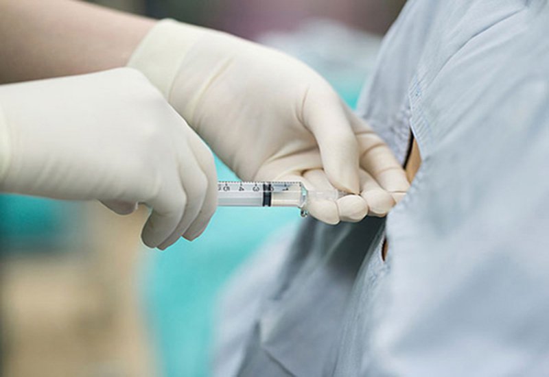
The principle of high frequency burning method is to convert alternating current into heat energy. A corrugated needle is inserted into the center of the tumor, an electric current is passed, generating heat to destroy the tumor. The electric current will be adjusted to ensure the generation of heat at 60-100 degrees Celsius. This is a temperature that can cause instant death of tumor cells. The time to perform radiofrequency ablation usually lasts from 8-12 minutes, depending on the specific case.

1.2. Cone-beam computed tomography in the treatment of liver tumors
New generation DSA angiograms can be used for computed tomography. The method of computed tomography right on the angiogram table is called cone-beam computed tomography (CBCT).
Cone-beam computed tomography will be done at two times, that is:
Taken just before starting the radiofrequency ablation machine to make sure the ablation needle is in the correct position in the tumor need intervention. Taken after radiofrequency ablation to determine the extent of necrosis and residual tumor. The method is absolutely contraindicated in patients with coagulopathy. For patients with central liver tumors, subcortical liver tumors adjacent to the gastrointestinal tract structures, patients with a history of biliary-enteric anastomosis,... Doctors must consider very carefully before performing the procedure.

2. Preparation before performing cone-beam computed tomography in radiofrequency ablation for liver tumors
On the patient's side, to prepare for the procedure, the patient will be consulted by medical staff about the condition of the patient. condition, carefully explain the procedure to coordinate with the implementation team. Patients need to fast for 6 hours before the procedure, can drink water but not more than 50ml.

3. Steps to perform cone-beam computed tomography in high-frequency ablation of liver tumors
4. Complications may be encountered when performing cone-beam computed tomography in radiofrequency ablation for liver tumors.
Abdominal bleeding: The patient will be closely monitored for hemodynamic factors. If you are anemic, you will be given blood transfusions and clotting factors. If medical treatment is not effective, the doctor will recommend surgery to stop the bleeding. Hollow visceral perforation: Usually monitored medically and treated with antibiotics. If that doesn't work, the patient will be indicated for surgery to stitch the perforation. A common late complication is biliary atresia. If the patient is not clinically symptomatic, intervention may not be necessary. If the patient has complications of biliary tract infection, jaundice, surgery must be performed to dilate and drain the biliary tract.
In order to meet the needs of medical examination and treatment, currently Vinmec International General Hospital has been and continues to bring in a system of modern machines such as magnetic resonance imaging (MRI), computed tomography (CT), X-ray, etc. .. into the work of medical examination and treatment, diagnostic imaging, disease treatment. With a full range of modern medical equipment, the implementation of high-frequency ablation technique for liver tumor treatment allows the doctor to control the entire procedure, avoiding maximum damage. Injury to blood vessels, nerves, trachea, esophagus should be very safe, and at the same time reduce the maximum tumor size.
Especially to bring high efficiency in disease examination and treatment, Vinmec now also designs many accompanying medical services such as the package of screening and early detection of liver cancer suitable for many at-risk subjects. Risk of various diseases including the liver cancer screening package - detecting cancer early when there are no symptoms brings convenience and satisfaction to all customers.
Để đặt lịch khám tại viện, Quý khách vui lòng bấm số HOTLINE hoặc đặt lịch trực tiếp TẠI ĐÂY. Tải và đặt lịch khám tự động trên ứng dụng MyVinmec để quản lý, theo dõi lịch và đặt hẹn mọi lúc mọi nơi ngay trên ứng dụng.
Dịch vụ từ Vinmec
-
 Nam giới u gan, vàng da điều trị như thế nào?
Bố của em đau rất nặng được chẩn đoán là u gan. Bố em ngày càng đau, sút cân, vàng da, mắt, vùng gan sưng lên thấy rõ, em rất lo sợ. Vậy bác sĩ cho em hỏi nam giới ...
Đọc thêm -
 Viêm lộ tuyến cổ tử cung nhẹ có đốt được bằng sóng cao tần RFA không?
Em năm nay 27 tuổi mới sinh em bé được 2 tháng rưỡi. Khi mang bầu 3 tháng, em đi khám phụ khoa phát hiện bị viêm lộ tuyến cấp độ 1. Em được kê đơn thuốc đặt và rửa ...
Đọc thêm -
 Phương pháp điều trị u gan đa ổ hiệu quả?
Bố em bị u gan đa ổ, huyết khối tĩnh mạch trái, khối u to 12m. Hiện tại, bố em đang nút mạch ở bệnh viện nhưng chưa thấy có hiệu quả. Bác sĩ tư vấn giúp em có phương ...
Đọc thêm -
 Điều trị bướu cổ tuyến giáp kích thước 18x24mm như thế nào?
Em có bướu cổ tuyến giáp 18x24mm. Em đang nuôi con nhỏ 5 tháng. Vậy bác sĩ cho em hỏi điều trị bướu cổ tuyến giáp kích thước 18x24mm như thế nào? Em có nên sử dụng đốt sóng cao ...
Đọc thêm -
 Bị viêm lộ tuyến cấp 1 có nên đốt sóng cao tần không?
Bác sĩ cho em hỏi em bị viêm lộ tuyến cấp 1 có nên đốt sóng cao tần không? Cảm ơn bác sĩ.
Đọc thêm