
Digital background removal and dilation, renal artery stenting
1. What is renal artery stenosis?
Renal artery stenosis may not cause any signs or symptoms until it is very advanced. However, renal artery stenosis can also be discovered incidentally during a routine physical exam, or your doctor may suspect the problem if you have unusually high blood pressure, such as sudden or worsening blood pressure. of unknown cause, early-onset hypertension before age 30 or after age 50. As renal artery stenosis progresses, other signs and symptoms may include high blood pressure that is difficult to treat, renal murmur, fluid overload, heart failure...
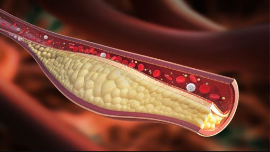
Two major causes of narrowing of the arteries kidney is atherosclerosis and muscular dysplasia. In addition, the renal artery can also be narrowed due to other less common conditions such as vasculitis, neurofibromatosis, or an intra-abdominal mass pressing on the renal artery. Regardless of the cause, when renal artery stenosis has reached a level that causes symptoms, in addition to medical treatment and risk factors control, renal artery revascularization is essential.
Current revascularization interventions to treat renal artery stenosis are renal artery stent dilation and renal artery bypass surgery.
2. What is a digital background eraser and angioplasty, renal artery stenting?
Thanks to the technique of fluoroscopy under the screen and the use of a sufficient amount of contrast medium, the renal artery system will be clearly visible and the location of the narrowing will be determined. From there, the doctor will approach the instrument, inflate the balloon and press the plaque against the vessel wall. As a result, the lumen of the vessel increases, and blood flow to the arteries improves. To maintain this result, a metal support (stent) will be saved. However, because it is a foreign material, there is a risk of embolism, after the intervention of placing the rack, the patient needs to take long-term antiplatelet drugs to stabilize the function of this device.
In summary, the clinical indications for digitized angiography and angioplasty, renal artery stenting are currently applied in cases where there is evidence of renal artery stenosis of more than 50% of all causes and symptoms. clinical symptoms such as hypertension, renal failure. At the same time, the patient does not have contraindications such as allergy to iodine contrast agents, severe renal failure, blood clotting disorders or pregnant women to avoid the risk of radiation exposure to the fetus. However, the above contraindications are relative, especially when renal function is severely impaired, severe hypertension is difficult to control and is responsible for renal artery stenosis.

3. Process of digital imaging to remove background and dilation, install renal artery support
Means of monitoring breathing, pulse, blood pressure, electrocardiogram, SpO2 are mounted on the patient and the indicators are displayed on the screen for easy observation. After cleaning the inguinal and genital areas, the doctor will cover the area with a sterile tissue to prepare for the intervention. In case the patient is overstimulated and poorly cooperated, it is necessary to consider appointing a sedative to facilitate the process.
At the intervention site, the doctor will administer local anesthesia on the right femoral artery. When the anesthetic takes effect, the doctor will insert a needle and insert the catheter into the artery. Under the background digitization and contrast screen, the doctor adjusted and guided the catheter to the location of the bifurcation from the abdominal aorta of the renal artery on the side of the injury to the intracranial arterial circulation. Cobra catheters are used to look for narrow passages that are indicated for intervention.
When accessing the location and determining the necessary features of the narrowed renal artery on the DSA, the doctor will proceed to place a catheter into the base of the damaged renal artery. Next, the microcatheter and microwire will be threaded through the narrowing, to the interlobar arteries. Next, the balloon is slowly inflated, and the lumen is widened. Then, when the ball is deflated, a metal support frame will be raised over the narrow spot, unfolding the corresponding bracket to the narrow position.
Finally, the doctor will determine whether the result is satisfactory or not based on the DSA image, the position of the bracket at the narrowed renal artery, the frame is completely open and the lumen is well re-opened. . If the plaque thickness is large, an incomplete narrowing of the lumen may be allowed with the degree of stenosis not exceeding 30%. In addition, the position of the lumen support has the distal end located at least 1cm below the occlusion site, the proximal end convex into the aortic lumen but not more than 3mm. At the same time, the doctor will also check the pulse before, during and after the recirculation is normal, there are no signs of thrombosis or dissection of the vessel wall. The same study was performed on selective angiography of the visceral body, superior mesenteric, and inferior mesenteric angiograms with a Cobra catheter (YASHIRO, liver, RH...)
When all of the above requirements are met, Interventional instruments will be withdrawn. The puncture artery site will be manually compressed for 15 minutes, fixed for 24 hours, or used with angioplasty and compression bandages within 6 hours. During this time, the patient is limited to bed movement.

4. Complications may be encountered in digitized scanning and dilatation, renal artery stenting
Arterial perforation Fracture displacement scaffolding Mucosal injury causing arterial dissection Abdominal bleeding Thromboembolism causing renal, mesenteric infarction Complications with anesthesia or contrast media Risk of radiation exposure during DSA .

In summary, when severe renal artery stenosis is symptomatic, revascularization is important, both to control hemodynamics and to preserve renal function. In which, digital scan to erase the background and dilation, placing renal artery bracket is the preferred method to choose compared to bypass surgery, with minimal intervention but still ensuring treatment efficiency.
Dr. Le Xuan Thiep has strengths in performing advanced and difficult magnetic resonance and computed tomography techniques such as: coronary computed tomography, cardiac function, cerebral magnetic resonance, perfusion brain and organs,..
If you notice any unusual health problems, you should visit and consult a specialist
Để đặt lịch khám tại viện, Quý khách vui lòng bấm số HOTLINE hoặc đặt lịch trực tiếp TẠI ĐÂY. Tải và đặt lịch khám tự động trên ứng dụng MyVinmec để quản lý, theo dõi lịch và đặt hẹn mọi lúc mọi nơi ngay trên ứng dụng.
Dịch vụ từ Vinmec
-
 Viêm động mạch Takayasu: Bệnh hiếm gặp, dễ bỏ sót
Viêm mạch takayasu là tình trạng viêm trong thành mạch máu, đôi khi gây ra tắc nghẽn hoàn toàn động mạch. Bệnh takayasu là một bệnh hiếm gặp, nguyên nhân chính xác chưa được biết rõ. Điều trị viêm mạch ...
Đọc thêm -
 Chụp động mạch thận chẩn đoán nhiều bệnh lý phức tạp
Chụp mạch máu thận hay còn gọi là chụp động mạch thận bằng cách sử dụng tia X chụp mạch máu ở thận khi tiêm chất cản quang vào mạch máu của thận. Kỹ thuật này được sử dụng để ...
Đọc thêm -
 Rò động-tĩnh mạch màng cứng tủy sống
Rò động tĩnh mạch màng cứng tủy sống thường khó chẩn đoán nhưng nếu được phát hiện kịp thời thì việc điều trị sẽ trở lên dễ dàng hơn. Bài viết hôm nay sẽ đi sâu vào các khía cạnh ...
Đọc thêm -
 Một số cách can thiệp động mạch thận
Can thiệp động mạch thận có chỉ định thực hiện để kiểm soát các triệu chứng của hẹp động mạch thận và ngăn ngừa các biến chứng. Những bệnh nhân thường được chỉ định can thiệp mạch thận là những ...
Đọc thêm -
 Quy trình chụp cộng hưởng từ tĩnh mạch không tiêm thuốc đối quang từ
Cộng hưởng từ tĩnh mạch không tiêm thuốc đối quang từ tham gia vào chẩn đoán và thực hiện theo dõi những diễn biến của bệnh lý liên quan đến tĩnh mạch như thông động tĩnh mạch, huyết khối, dị ...
Đọc thêm