
Digital imaging to erase the background and remove atherosclerotic plaques to treat arterial stenosis
The article is expertly consulted by Specialist Doctor I Tran Cong Trinh - Radiologist - Radiology Department - Vinmec Central Park International General Hospital and Master, Doctor Le Xuan Thiep - Doctor of Radiology - Department of Diagnostic Imaging - Vinmec Ha Long International General Hospital.
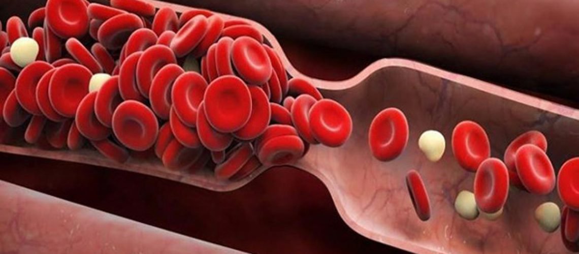
1. What is artery stenosis?
The main cause of limb artery stenosis is atherosclerosis of the limb arteries. Risk factors for atherosclerosis include:
Age 55-60 and gender; Smoke; Diseases such as: Diabetes, hypertension, dyslipidemia, hyperhomocysteinemia. Recognizing symptoms:
There is a feeling of muscle spasm when exerting or walking a long distance. The pain goes away with rest. However, over time, pain will appear even when the patient is at rest. Pain in the buttocks or thighs due to damage to the iliac arteries; pain in the calf due to damage to the femoral-popliteal artery; pain in the feet due to damage to the arteries of the legs. The feeling of burning pain or numbness, cold extremities, is relieved if you let your legs fall or stand up. Muscular dystrophy, hair loss, thick nails. Skin examination revealed green, cold skin, with small ulcers, well-defined, on the arterial blood supply. Physical examination reveals a weak pulse or, worse, loss of pulse. To treat occlusive stenosis of the extremities, angioplasty and percutaneous stenting are the basic techniques. However, in many cases it is necessary to digitize the background and remove atherosclerotic plaques and chronic thrombosis before angioplasty or stenting.
This method uses specialized instruments to dredge and remove plaque caused by arterial occlusion, chronic thrombotic plaques clinging to the wall of the vessel to widen the lumen for other techniques.
2. Indications and contraindications for digital imaging to erase the background and remove atheroma for treatment of arterial occlusion

Re-stenosis in stent 2 Chronic occlusive stenosis of the arteries of the extremities due to atheroma, thrombosis Atherosclerotic plaque treatment is contraindicated in the following cases:
Renal failure grade IV Coagulation disorders and loss of control Pregnant women Diffuse arterial damage Allergy to iodine contrast agents
3. Steps to perform digital scan to erase the background and remove atheroma to treat narrow artery occlusion
Step 2: Ask the patient to lie on his or her back on the x-ray table and then place an intravenous line and local anesthetic, pre-anesthetic can be injected in exceptional cases such as young children.
Step 3: Place the tube in the vessel. Depending on the location and purpose of the intervention, it is possible to open the way into the downstream or upstream vessels.
Step 4: Conduct angiography of the lower extremity vasculature through the catheter and then evaluate the entire lower extremity vascular system. Then use the catheter, guide and micro-catheter, micro-conductor to pass through the narrow - occluded site and apply intravascular techniques.
Step 5: Insert the atheroma removal tool into the narrow position through the wire. Then, activate the electrical switch to perform atheroma dredging. Repeat this operation until the lumen is recanalized. 820
Step 6: Combine with traditional recanalization techniques and angiography to assess circulation after recanalization.
Step 7: Withdraw the tube into the vessel, then apply pressure, close the path into the vessel to end the procedure.
4. Handling accidents
Before taking a job at Vinmec Central Park International General Hospital, the position of Doctor of Radiology from September 2017, Doctor Tran Cong Trinh worked at Gia Dinh People's Hospital since 2007. -2017. In his role, Doctor Tran Cong Trinh has participated in guiding the teaching of students, residents, specialists and new doctors entering the department. Advanced and difficult magnetic resonance and computed tomography techniques such as: coronary computed tomography, cardiac function, cerebral magnetic resonance, cerebral perfusion and visceral organs, ..
If problems are found Abnormal health should be examined and consulted with specialists
Để đặt lịch khám tại viện, Quý khách vui lòng bấm số HOTLINE hoặc đặt lịch trực tiếp TẠI ĐÂY. Tải và đặt lịch khám tự động trên ứng dụng MyVinmec để quản lý, theo dõi lịch và đặt hẹn mọi lúc mọi nơi ngay trên ứng dụng.
LEARN MORE
Application of digital background angiography (DSA) in cardiovascular Causes of coronary atherosclerosis Coronary angiography - Dual effects in diagnosis and treatment of coronary artery disease
Dịch vụ từ Vinmec
-
 Có thể dùng thuốc cản quang chụp mạch máu để chụp ống tiêu hóa không?
Bác sĩ cho em hỏi có thể dùng thuốc cản quang chụp mạch máu để chụp ống tiêu hóa không? Cảm ơn bác sĩ tư vấn.
Đọc thêm -
 Chụp số hóa xóa nền và điều trị lấy huyết khối cấp tính động mạch chi
Phương pháp chụp số hóa xóa nền (DSA) hút huyết khối qua ống thông được thực hiện bằng cách đưa ống thông có đường kính phù hợp vào tới vị trí cục huyết khối, sau đó tiến hành hút áp ...
Đọc thêm -
 Người bị sốc phản vệ thuốc cản quang có khả năng sốc phản vệ thuốc tê không?
Bác sĩ cho em hỏi người bị sốc phản vệ thuốc cản quang có khả năng sốc phản vệ thuốc tê không? Em cảm ơn bác sĩ.
Đọc thêm -
 Chụp cắt lớp vi tính có phải sử dụng thuốc cản quang không?
Chào bác sĩ, Bác sĩ cho em hỏi chụp cắt lớp vi tính có phải sử dụng thuốc cản quang không? Khi nào chụp cắt lớp vi tính có thêm chỉ định sử dụng thuốc cản quang không? Em cảm ...
Đọc thêm -
 Vinmec có kỹ thuật nào chẩn đoán u tủy không?
Em muốn đi chụp ống tủy xem có u tủy không nhưng không biết chụp thế nào? Ở Vinmec có kỹ thuật nào chẩn đoán u tủy không? Cảm ơn bác sĩ tư vấn.
Đọc thêm