
Emergency ophthalmoscopy
1. What is ophthalmoscopy?
Ophthalmoscopy helps detect diseases of vitreous (transparent environment of the fundus), retina, optic nerve (optic nerve),... Early detection of lesions helps doctors monitor and manage timely treatment, thereby reducing the risk of complications that damage the patient's vision.
2. When is ophthalmoscopy needed?
Damage to the optic nerve Retinal tear or detachment Glaucoma ( Glaucom) Loss of sharp vision due to aging (macular degeneration). Retinitis Hypertension Diabetes mellitus Melanoma skin cancer

3. When is emergency ophthalmoscopy performed?
Ophthalmoscopy is indicated in all for emergency fundus examination in the intensive care unit or emergency bed, it is necessary to observe the condition of the retina, optic disc, and macula, with or without damage to the fundus. related to systemic emergency diseases such as: poisoning with chemicals, drugs, alcohol, asphyxiation, drowning, infectious diseases causing fundus hemorrhage....
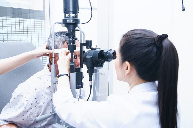
4. Ophthalmoscopy methods?
Direct ophthalmoscopy: usually performed in a dark room. The patient will be asked to remove contact lenses (if present), keep their eyes looking straight ahead, and keep their head still. The doctor will shine the light directly into the patient's eyes. Direct ophthalmoscopy for virtual and lateral images, magnifying the image 10-15 times. Observation of the eye area is limited (10 - 15 degrees) and difficult to see if the environment is cloudy. This procedure can be done with or without dilated pupils. Indirect ophthalmoscopy, also known as inverted ophthalmoscopy. The indirect ophthalmoscope is worn over the doctor's head and the patient is asked to lie down or sit in a prone position. The indirect ophthalmoscopy method gives real and inverted virtual images, magnifying the image 2 - 5 times. This is a difficult method to perform and requires specialized tools and higher skills. However, this method has advantages over direct ophthalmoscopy, because the doctor can see better in the eye if there is a cataract. At the same time, a 3-D image of the back of the fundus can be viewed to help the doctor identify certain eye conditions such as growths, optic nerve edema, or if the retina is detached. In addition, indirect ophthalmoscopy also helps the doctor see a larger area (160 degrees) than direct ophthalmoscopy.
5. How is ophthalmoscopy performed?
5.2. Ophthalmoscopy is usually done in 5-10 minutes according to the following steps:
The doctor faces the patient, the doctor's right eye examines the patient's right eye or vice versa when looking at left eye fundus. The doctor observes the patient's retinal condition, the patient's optic disc, the patient's macula, and assesses the condition of related damage. In cases where the patient's retina cannot be seen, adjust the lens system on the ophthalmoscope to a diverging or converging lens to observe the patient's fundus image. The scope of the fundus being observed is very narrow, less than 1mm wide, so the illuminator must be placed very close to the patient and moved around the areas of the fundus. For young patients, uncoordinated can be anesthetized the surface of the eyeball and use the rim of the eyelids for ophthalmoscopy. Fundus dilation is a fairly safe procedure, with patients having almost no complications. In rare cases, the patient may experience discomfort from the eye drops or from the light of the lamp. If there is a side effect of mydriasis, the eyeball surface should be numbed, immediately stop applying the drug, wash the eyes with physiological saline. The doctor will carefully monitor the patient's progress to have appropriate treatment.
Eye Specialist - Vinmec International General Hospital always receives and handles patients who are having eye problems. With a system of modern equipment and a team of experienced doctors, they will directly examine and advise on the best treatment for the current condition. All procedures are carried out in a methodical and intensive manner, so customers can be assured of medical services at Vinmec.
Để đặt lịch khám tại viện, Quý khách vui lòng bấm số HOTLINE hoặc đặt lịch trực tiếp TẠI ĐÂY. Tải và đặt lịch khám tự động trên ứng dụng MyVinmec để quản lý, theo dõi lịch và đặt hẹn mọi lúc mọi nơi ngay trên ứng dụng.
Dịch vụ từ Vinmec
-
 Tác dụng của thuốc Brinzolamide
Brinzolamide được biết đến với các tên phổ biến khác như Azopt, Befardin, là một chất ức chế anhydrase carbonic được sử dụng để hạ nhãn áp ở những bệnh nhân bị bệnh tăng nhãn áp góc mở hoặc tăng ...
Đọc thêm -
 Mắt nhìn mờ đục, đau khi ánh sáng chiếu vào sau khi bóng đập vào mắt có sao không?
Cháu đá bóng bị đập vào mắt, mắt cháu ngoài bị bầm. Khi nhìn, cháu còn có hiện tượng bị mờ đục, nhìn ra rất nhiều vằn đen và bị đau mắt khi ánh sáng chiếu vào. Vậy bác sĩ ...
Đọc thêm -
 Ai cần thận trọng khi sử dụng thuốc tăng nhãn áp?
Sử dụng thuốc tăng nhãn áp cần phải theo đúng chỉ định của bác sĩ chuyên khoa bởi không phải trường hợp bệnh nhân nào cũng có thể dùng thuốc này. Vậy ai cần thận trọng khi sử dụng thuốc ...
Đọc thêm -
 Helicobacter và bệnh tăng nhãn áp
Bệnh tăng nhãn áp ảnh hưởng đến hơn 70 triệu người trên toàn thế giới. Bệnh tăng nhãn áp là một bệnh lý thần kinh thị giác tiến triển được đặc trưng bởi sự thay đổi của đầu dây thần ...
Đọc thêm -
 Mắt giật theo nhịp kèm hoa mắt là dấu hiệu bệnh gì?
Em bi hoa mắt, nhìn thấy chớp sáng, đôi khi mắt mất thị lực hay bị giật theo nhịp. Vậy bác sĩ cho em hỏi mắt giật theo nhịp kèm hoa mắt là dấu hiệu bệnh gì? Em cảm ơn ...
Đọc thêm