
Endoscopic marrow stimulation drill
1. What is knee cartilage?
The articular cartilage is mainly made up of fluid, they help absorb most of the forces acting on the 2 ends of the bones. This ensures continuous movement of the knee joint during human activities.
2. Common injuries to knee cartilage
Cartilage can also be damaged by trauma or degenerate during daily activities. However, articular cartilage cannot be able to restore itself, but these injuries will get worse day by day, leading to a complete loss of articular cartilage and exposing bone under the cartilage.
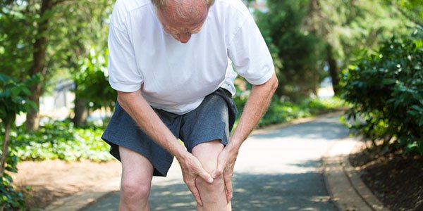
When the cartilage layer is lost, the joints will lose their smooth surface with the ability to absorb force during movement. Therefore, the patient will have swelling and pain in the knee joint when moving, the pain may be worse when squatting or walking up the stairs. Patients can also get stuck in the knee joint because of the detachment of cartilage in the knee joint, which, if not treated promptly, can lead to limited muscle movement and muscle atrophy.
Cartilage damage can be caused by mechanical damage such as:
Knee cruciate ligament rupture Inflammatory synovitis Meniscus tear Diseases that can cause cartilage damage:
Decompressive cartilage inflammation Rheumatoid polyarthritis Rheumatoid arthritis Levels of cartilage damage include:
Cartilage damage is divided into many levels, each type of damage with different levels will be indicated for treatment by different methods.
Grade I: articular cartilage softening Grade II: fibrinoid scarring and superficial fissures on the surface of articular cartilage Grade III: Deep fissures in the cartilage surface but no bone is exposed Grade IV: Loss of articular cartilage, exposing subchondral bone

3. Some current knee osteoarthritis treatment surgery
3.1 Laparoscopic surgery to wash the knee joint
Basically, this surgery washes the knee joint using physiological saline solution to wash through 2 trocar tubes. Theoretically, this method can remove small pieces of foreign bodies that appear due to the process of cartilage erosion, and remove cytokines.
Arthroscopic surgery to wash the joint, remove the bursitis tissue, remove the foreign body in the knee joint, remove the marginal bone spur. The pieces of articular cartilage that are still attached but have lost their firmness and are in danger of detaching and breaking, creating foreign bodies are also removed. Meniscus tears due to degeneration will also be treated.
3.2 Shaft repair bone chisels
The main purpose of this method is to change the mechanical axis of the leg, shifting the weight bearing from the degenerative joint cavity to the healthy space based on the physiological axis. This helps to reduce the pressure on the degenerative joint surface, slows down the degenerative process and helps patients reduce pain.
This method is usually indicated for cases of early knee osteoarthritis, 1-cavity degeneration, often seen in patients with leg deformities in the form of internal and external curvature (O, X or K type). Can choose the location of chisels in the tibial plateau or on the femoral condyle, the "V closed" or "Open V" type can be chiseled depending on the case.
3.3 Endoscopic technique of drilling for pulp stimulation
Endoscopic technique of marrow stimulation drilling is a technique applied in the treatment of knee cartilage damage.
The cartilage damage has 4 different levels, but this technique only applies to grade III and IV damage, when the articular cartilage has shown signs of severe damage, it is necessary to regenerate new cartilage. Not only that, this technique is only indicated for lesions from 2 to 3cm2. Cases with lesion area > 3cm2 or diffuse degenerative lesions do not use this method.
3.3.1 Preparation before surgery
Performer: The person performing this surgery must be a specialist in orthopedic surgery and have been fully trained in arthroscopic knee surgery. Instruments and tools: Arthroscopy machine, knee arthroscopy instruments and instruments to close the marrow stimulation. Patients should be tested, explained about surgical methods, risks and rehabilitation exercises after surgery. The patient cleans his body and fasts for 6 hours before surgery.

3.3.2 How is the endoscopic technique of drilling for pulp stimulation?
In endoscopic spinal stimulation, the doctor will diagnose through the endoscope to assess the size and depth of the lesion by maximally flexing and extending the entire facet of the joint. From there, evaluate and treat the associated lesions.
Resection of loose cartilage and damaged cartilage edges until the cartilage is normal. Remove the bony shoots and then scrape off the callus at the base of the affected area. This increases the adhesion of blood clots, improving the nutrients that feed the cartilage. Use a closing tool to create holes with a distance of 3-4mm to stimulate the pulp in the damaged area. Stop the water pump and determine the blood flow from the bone marrow to the holes just created. Do not drain the joint and suture the incision. After surgery, some studies also show that the newly formed cartilage area is mainly fibrocartilage with the composition of basic substances, and the chondrocytes are also sparse. Therefore, this method is often combined with autologous chondrocyte transplantation or autologous or allogeneic bone grafting.
3.3.3 Monitoring and handling of accidents
Patients use prophylactic antibiotics before surgery and 24 hours after surgery. After surgery, use pain relievers and cold compresses.
Exercise mode: Take crutches and gently touch the sore leg for 6-8 weeks. Practice passive bending and stretching for the first 4-6 weeks. Possible complications are arthritis or infection.
Endoscopic marrow stimulation drilling is a difficult technique, which needs to be performed at prestigious and highly specialized hospitals. Vinmec International General Hospital is one of the medical facilities with a team of doctors and nurses with many years of experience and a system of modern equipment, which is definitely a reliable address if you are in need of internal surgery. marrow stimulation drill.
Any questions that need to be answered by a specialist doctor as well as customers wishing to be examined and treated at Vinmec International General Hospital, you can contact Vinmec Health System nationwide or register online HERE.
Dịch vụ từ Vinmec
-
 Giảm sưng khớp gối cho người cao tuổi bị viêm khớp thế nào?
Mẹ mình năm nay 84 tuổi. Trước đây mẹ mình từng bị bệnh thần kinh tọa chân nên di chuyển và đi lại hơi khó khăn. Khoảng 2 tháng gần đây bà có biểu hiện sưng khớp gối và sưng ...
Đọc thêm -
 Điều trị tụ dịch trong bao sụn khớp gối như thế nào?
Mỗi khi ngồi gấp 2 đầu gối là tôi bị đau, phải co duỗi từ từ mới đứng dậy được. Tôi đi khám, chụp MRI khớp gối và được chẩn đoán tụ dịch trong bao sụn khớp gối và bao ...
Đọc thêm -
 Tổn thương dây chẳng khớp gối có sao không?
Em bị vấn đề về chân. Chân em chơi đá banh lúc nhỏ bị đau nhưng thời gian cảm thấy không sao nhưng giờ gối em bị yếu và lỏng! Như vậy gối em bị gì thưa bác sĩ?
Đọc thêm -
 Khớp gối lỏng lẻo sau chấn thương phải làm sao?
Dạ thưa bác sĩ, hồi trước em có đá banh và gặp phải chấn thương ở đầu gối, lúc đó đầu gối rất đau và khó di chuyển nhưng bây giờ em lại hay bị trật khớp ở vùng bị ...
Đọc thêm -
 Có thể phẫu thuật thay lại khớp gối ở Vinmec ngay không?
 Mẹ em đã thay khớp gối 12 năm. Hiện mẹ em đang rất đau nhức và có chỉ định thay khớp mới nhưng bác sĩ báo chưa có hàng. Cho em hỏi bệnh viện mình đang có những loại khớp ...
Đọc thêm