
Learn about anal fistula magnetic resonance imaging
1. What is anal fistula?
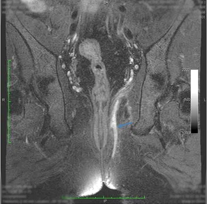
Anal fistula is an anorectal disease that manifests as an abnormal tunnel between the anal canal and the perianal skin.
Anal fistula originates from an anal gland located between the internal and external anal sphincters and drains into the anal canal. When the gland's outlet is blocked, a perianal abscess is formed and can spread to the surface of the skin. Improper treatment, improper technique or untreated perianal abscesses will cause anal fistula.
Based on the internal and external sphincter, anal fistula can be divided into 4 types (according to the Park classification) or 5 grades (according to the Garg classification).

2. How is an anal fistula magnetic resonance imaging?
2.1 Preparation The anal fistula magnetic resonance imaging method adheres to some general principles of magnetic resonance imaging, that is, the removal of metal objects/devices in the body. You will be asked to be sure not to wear metal devices after surgery or implantation of electronic devices that are affected by strong magnetic fields.
Magnetic resonance anal fistula includes two phases before drug injection and after injection of magnetic contrast (magnetic contrast). The main purposes of using contrast agents are to help visualize the fistula and evaluate the tissue surrounding the fistula. Although allergic reactions are rare with contrast agents, your doctor will ask you about your medical history and advise you on the benefits and risks of the drug in each individual case.
Sedatives or anesthetics may be administered intravenously in special cases. At this time, you may have to fast for about 4-6 hours before to ensure safety during anesthesia or sedation.
2.2 Perform An anal fistula MR scan about 30-45 minutes. You will lie on the imaging bed and the technician will place a special device called a “Coil” around your abdomen. The scan table will be inserted into the “cage” and the technician will begin to take pictures of the pelvis, including the anus and the area around the anus. You will be given an injection by the technician between the scans. After the injection, the anal fistula will be more prominent on the image, helping the doctor to better identify and evaluate the lesion.
This shooting process will cause certain noise, and you will feel some heat in the shooting area. If you are sedated or sedated, you will be monitored for hygiene signs such as heart rate, breathing rate, and blood oxygen levels during the scan through devices that are captured on your body.

3. Advantages and disadvantages of anal fistula magnetic resonance imaging?
Besides the means of X-ray, CT Scan, colonoscopy, anal ultrasound ... magnetic resonance is an advanced means to evaluate and classify anal fistula. Magnetic resonance has many advantages in accurately describing the perianal anatomy, assessing the relationship between the fistula and the pelvic diaphragm (lift muscle) and the rectal fossa. This is an important part of helping surgeons before surgery.
However, the disadvantage of anal fistula is that it takes a long time and is expensive, so it is difficult to reach the masses. In addition, it is difficult or impossible for people with claustrophobia to perform. this technique.
4. What results did you get after anal fistula magnetic resonance imaging?
The radiologist will analyze and give results based on images taken through each thin slice of the pelvis. Thereby, your fistula will be classified according to the existing classification table. Simultaneously, the parenchyma surrounding the fistula will be evaluated and noted for noticeable abnormalities, including complications of the fistula (eg, infection, abscess, recurrent fistula...).
The surgeon will base on the results of magnetic resonance imaging and other necessary test results to evaluate and thereby select the appropriate fistula treatment method.
5. Equipment for anal fistula magnetic resonance imaging?
For anal fistula magnetic resonance imaging, the best hospital should use a machine of 1.5 Tesla or more. The GE 3 Tesla magnetic resonance machine used at Vinmec Nha Trang is capable of performing this technique well with the appropriate protocol.
Vinmec International General Hospital is one of the hospitals that not only ensures professional quality with a team of leading medical doctors, modern equipment and technology, but also stands out for its examination and consultation services. comprehensive and professional medical consultation and treatment; civilized, polite and safe medical examination and treatment space.
Customers can go directly to Vinmec Nha Trang to visit or contact hotline 0258 3900 168 for support.
Dịch vụ từ Vinmec
-
 Chụp cộng hưởng từ rò hậu môn khi nào?
Rò hậu môn là bệnh lý gây sụt giảm chất lượng cuộc sống của người bệnh vì gây đau đớn và thường xuyên tái phát. Bệnh cần được điều trị triệt để bằng phương pháp phẫu thuật. Chụp cộng hưởng ...
Đọc thêm -
 Tổn thương đường tiêu hóa của bệnh Crohn
Bệnh Crohn là một thể của bệnh lý viêm ruột IBD (Inflammatory Bowel Disease) gây ra viêm nhiễm, ăn sâu vào thành của đường tiêu hóa gây loét, chảy máu. Bệnh gặp ở mọi lứa tuổi nhưng chiếm tỷ lệ ...
Đọc thêm -
 Sưng vết mổ áp xe hậu môn có bất thường không?
Cách đây tầm 7 tháng, con có mổ áp xe hậu môn. Hôm nay, con sờ lại thấy chỗ từng mổ áp xe sưng lên nhưng con chưa thấy đau. Bác sĩ cho con hỏi, sưng vết mổ áp xe ...
Đọc thêm -
 Đau nhức quanh hậu môn kèm theo táo bón là dấu hiệu bệnh gì?
Chào bác sĩ. Tôi 60 tuổi nữ, đầu tiên tôi bị nhức hậu môn ngứa, điều trị xoa thuốc hết nhức và hết ngứa, tiếp sau đó táo bón và sưng một bên hậu môn. Tôi có ngâm phèn 2 ...
Đọc thêm -
 Trẻ sơ sinh bị rò hậu môn điều trị như thế nào?
Bé nhà em 2 tháng tuổi bị lỗ rò hậu môn, lên mủ. Bé uống thuốc xong rồi lại hết rồi lại lên. Ổ mủ không to, không sưng đau, bé không quấy khóc, không sốt. Vậy bác sĩ cho ...
Đọc thêm