
Pelvic magnetic resonance imaging procedure without injection of magnetic contrast
1. Outline
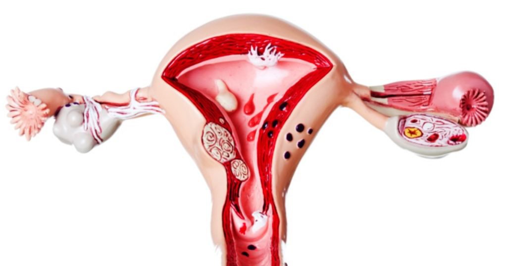
When a suspected pelvic lesion is detected after initial imaging, magnetic resonance imaging may be indicated in these cases for the purpose of surveying the pelvic organ and lesion.
Pelvic magnetic resonance imaging provides clear, high-resolution and good-contrast images that improve patient diagnosis and prognosis in treatment.
2. Indications for pelvic magnetic resonance imaging without injecting magnetic contrast
3. Contraindications to pelvic magnetic resonance imaging without contrast injection
4. Preparation for pelvic magnetic resonance imaging without contrast injection
5. Steps to conduct pelvic magnetic resonance imaging without injecting magnetic contrast
5.2. Selection of pulse sequences for non-contrast pelvic magnetic resonance imaging from the initial localization; Pulse sequence 1: Horizontal T2W with fat removal, slice thickness 6cm, interval between slices 10% of slice thickness (0.6 mm or coefficient 1.0-1.1), L-R code scale,, FOV < 200mm , the cross-sectional saturation plate is placed above the slice to prevent vascular interference; horizontal plane against abdominal wall fat interference. Pulse sequence 2: T1W cross-sectional plane, fat removal, position of slices similar to sequence 1. Pulse sequence 3: T2W with fat removal, horizontal plane or subframe position, thickness 4-6 mm, spaced 10 % of the thickness of the cut, the plate is saturated in the horizontal plane against vascular interference. Pulse sequence 4: T2W in vertical plane, thickness 5mm, interval 10% of cut thickness (0 – 0.5mm). Note: it is possible to combine drugs to help reduce intestinal motility, instruct the patient to breathe to reduce the noise caused by breathing.
6. Read results of pelvic magnetic resonance imaging without contrast injection

Especially, Vinmec International General Hospital is the first unit in Southeast Asia to put into use the new 3.0 Tesla Silent Resonance Imaging machine from the US manufacturer GE Healthcare.
The machine currently applies the safest and most accurate magnetic resonance imaging technology available today, without using X-rays, non-invasive. Silent technology is very beneficial for patients who are young children, the elderly, patients with weak health or have just had surgery.
Để đặt lịch khám tại viện, Quý khách vui lòng bấm số HOTLINE hoặc đặt lịch trực tiếp TẠI ĐÂY. Tải và đặt lịch khám tự động trên ứng dụng MyVinmec để quản lý, theo dõi lịch và đặt hẹn mọi lúc mọi nơi ngay trên ứng dụng.
Dịch vụ từ Vinmec
-
 Quy trình chụp cộng hưởng từ tuyến tiền liệt có tiêm thuốc đối quang từ
Chụp cộng hưởng từ tuyến tiền liệt là phương pháp được sử dụng để chẩn đoán cho các trường hợp nghi ngờ ung thư tuyến tiền liệt hoặc theo dõi quá trình điều trị bệnh. Chụp MRI tuyến tiền liệt ...
Đọc thêm -
 Quy trình chụp cộng hưởng từ tĩnh mạch không tiêm thuốc đối quang từ
Cộng hưởng từ tĩnh mạch không tiêm thuốc đối quang từ tham gia vào chẩn đoán và thực hiện theo dõi những diễn biến của bệnh lý liên quan đến tĩnh mạch như thông động tĩnh mạch, huyết khối, dị ...
Đọc thêm -
 Quy trình chụp cộng hưởng từ cột sống ngực có tiêm thuốc đối quang từ
Chụp cộng hưởng từ cột sống ngực có tiêm thuốc đối quang từ là một phương tiện chẩn đoán hình ảnh hiện đại và hữu ích, cung cấp những hình ảnh rõ ràng về các cấu trúc giải phẫu tại ...
Đọc thêm -
 Quy trình chụp cộng hưởng từ tuyến vú có tiêm thuốc đối quang từ
Khối u tại tuyến vú là tình trạng thường gặp phải ở phụ nữ trong độ tuổi sinh đẻ. Sau khi phát hiện tổn thương tại tuyến vú, bệnh nhân sẽ được chỉ định các xét nghiệm cận lâm sàng ...
Đọc thêm -
 Quy trình chụp cộng hưởng từ động mạch chi trên có tiêm thuốc đối quang từ
Chụp MRI động mạch chi trên có tiêm thuốc đối quang từ thường đem lại hình chụp với độ tương phản cao, chi tiết giải phẫu tốt cho phép nhận diện các tổn thương về mặt hình thái và chức ...
Đọc thêm