
Retinal vitreous: What you need to know
1. Retina and some retinal lesions
The central area of the retina, called the macula, contains a high density of color-sensitive (light-sensing) photoreceptors. These cells, called cones, produce the sharpest visual images and are responsible for central and color vision. The peripheral area of the retina, which surrounds the macula, contains light-sensitive cells called rods, which respond to lower light intensities but are not sensitive to color. They are responsible for peripheral vision and night vision.
The retina is located near the optic nerve. The retina processes the information gathered by the photoreceptors and sends this information to the brain via the optic nerve. The optic nerve and retina are profusely supplied with blood by a rich vascular system. Part of this blood vessel supply comes from the choroid, which is the layer of blood vessels that lies between the retina and the outer white layer of the eye called the sclera. The central retinal artery (the retina's other major blood source) reaches the retina near the optic nerve and then branches out into the retina. Blood flows from the retina into the branches of the central retinal vein. The central retinal vein exits the eye in the optic nerve.
When examining a person's retina, a doctor will put drops in the eye to dilate the pupil. This allows the retina to be seen in much greater detail by ophthalmoscopy (which shines light through a magnifying lens into the back of the eye).
Because of the important role the retina plays in vision, damage to the retina can cause permanent blindness. Conditions such as retinal detachment, in which the retina is abnormally separated from its normal position, can prevent the retina from receiving or processing light. This prevents the brain from receiving information, thus leading to blindness. Retinal disorders are usually diagnosed and treated by an ophthalmologist. An ophthalmologist is a medical doctor who specializes in the evaluation and treatment of all types of eye disorders.

2. What is glass translation?
When we are young, vitreous is mainly a liquid gel. As we age and the eyes become nearsighted, this gel will gradually become more liquid. Recent research has suggested many similarities between the vitreous and the joints in our bodies (vitreous vitreous is similar to synovial fluid), which promises an opportunity to advance our understanding of this unique gel. this by learning more about joints in the body.
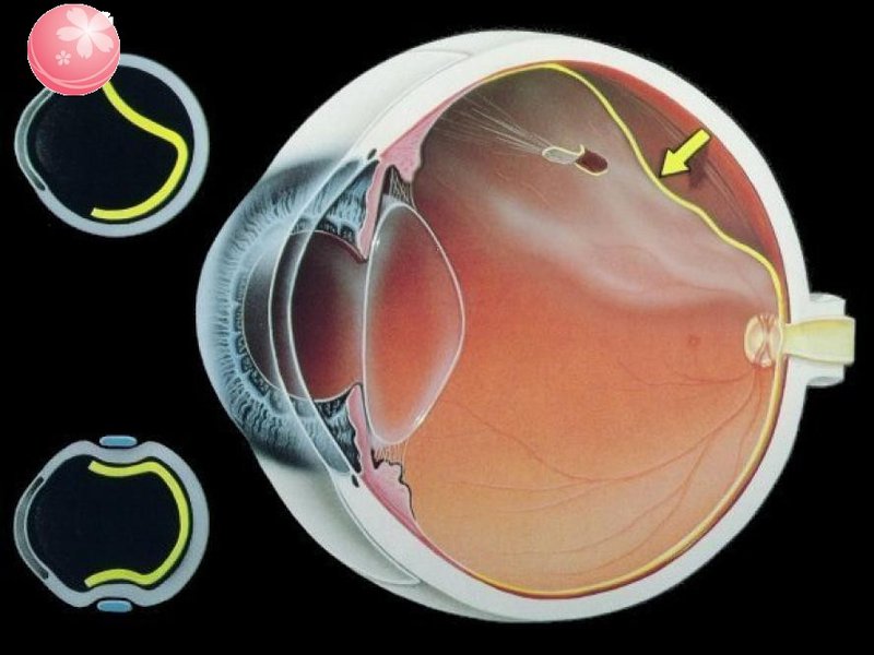
The back of the vitreous is adjacent to the retina and is attached to the retina at specific points. The retina is the light-receiving tissue inside the eye. The vitreous and retinal are most tightly adhered to the “vitreous base”. It is located in the most anterior part of the retina, i.e. the area immediately behind the iris. In most cases, the retina and vitreous do not separate even after posterior vitreous detachment (PVD). Here, a retinal tear can sometimes occur, leading to retinal detachment and blindness.
The main function of the vitreous is to keep the center of the eye transparent so that light can reach the retina and begin to have vision. The gel and its liquid allow oxygen and nutrients to flow from the front of the eye to the back of the eye. In adolescence, vitreous is a shock absorber during eye movements, head movements and strenuous activities of the body. It is also rich in antioxidants that help protect different parts of the eye. Today, vitreous is also seen as a reservoir for drugs that we inject into the eye to treat various diseases related to the macula and retina. For example, ophthalmologists often inject anti-VEGF and various steroids to treat macular degeneration or diabetic retinopathy in some patients.
In the advanced stages of diabetic retinopathy, blood can bleed into the vitreous causing vision loss due to physically obstructed light and irritation of the retina. A more common complication of diabetic retinopathy, known as macular edema, is also vitreous-related. In a patient with diabetic retinopathy, normal retinal vessels tend to leak over time. This leak can build up in the macula causing mild to severe vision loss.

Eye Specialist - Vinmec International General Hospital always receives and handles patients who are having eye problems. With a system of modern equipment and a team of experienced doctors, they will directly examine and advise on the best treatment for the current condition. All procedures are carried out in a methodical and intensive manner, so customers can be assured of medical services at Vinmec.
Để đặt lịch khám tại viện, Quý khách vui lòng bấm số HOTLINE hoặc đặt lịch trực tiếp TẠI ĐÂY. Tải và đặt lịch khám tự động trên ứng dụng MyVinmec để quản lý, theo dõi lịch và đặt hẹn mọi lúc mọi nơi ngay trên ứng dụng.
Dịch vụ từ Vinmec
-
 Các tác nhân gây oxy hóa trong cơ thể
Thực tế, có nhiều tác nhân gây oxy hoá trong cơ thể, chẳng hạn như khói bụi, ô nhiễm môi trường, thuốc lá, hoá chất, đường huyết cao,... Những yếu tố này có thể gây mất cân bằng oxy hoá ...
Đọc thêm -
 Phẫu thuật tán nhuyễn thủy tinh thể
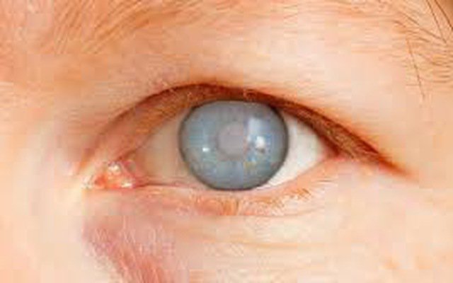
Nếu bạn trên 60 tuổi và thị lực bị mờ hoặc nhìn thấy các chấm đục, bạn có thể đã bị đục thủy tinh thể. Đây là một bệnh lý phổ biến ở người lớn tuổi và có thể được ...
Đọc thêm -
 Vai trò của axit mật trong bệnh xơ gan do hệ vi sinh vật đường ruột làm trung gian
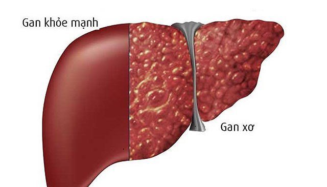
Các nghiên cứu trước đây đã chỉ ra rằng những thay đổi hệ vi sinh vật đường ruột hoặc rối loạn vi khuẩn ở bệnh nhân bị bệnh gan mãn tính hoặc xơ gan thường đi kèm với sự giảm ...
Đọc thêm -
 Mắt trẻ bong võng mạc điều trị như thế nào?
Bé nhà em mắt bị bong võng mạc. Vậy bác sĩ cho em hỏi mắt trẻ bong võng mạc điều trị như thế nào? Em cảm ơn bác sĩ.
Đọc thêm -
 Tạm biệt đục thủy tinh thể - mắt sáng khỏe, ông bà vui vẻ bên con cháu
Bước vào tuổi xế chiều, niềm mong ước lớn nhất của cha mẹ có lẽ là một sức khỏe tốt và một đôi mắt tinh anh để chứng kiến sự trưởng thành của con cháu. Thế nhưng theo thời gian, ...
Đọc thêm