
Signs that indicate you need a thyroid ultrasound
The article is professionally consulted by Master, Doctor Nguyen Thi Ngoc - General Internal Medicine - Endocrinology - Department of Examination & Internal Medicine - Vinmec Central Park International General Hospital.
1. What is a thyroid ultrasound?
Thyroid ultrasound is an examination method to visualize the thyroid structure by ultrasound to examine the normal thyroid gland as well as detect lesions in the thyroid gland, and at the same time survey the parathyroid gland lesions and other groups. involved nodes, if any.
Thyroid tissue elasticity ultrasound is an ultrasound technique that evaluates the stiffness of the tissue through the degree of elasticity of the tissue when subjected to mechanical force, a new method in the early diagnosis of thyroid cancer. Pathological tissues with the same echogenicity, on B-mode ultrasound images, it is difficult to distinguish benign or malignant properties, Doppler ultrasound can further support the diagnosis, malignant tumors often proliferate blood vessels. Therefore, elastography of thyroid tissue will help to add more information about the characteristics of the damaged tissue to increase the diagnostic ability.

2. Indications for thyroid ultrasound
According to Dr. Ngoc, this section is structured as follows:
- Detects solitary nodule, multinodular goiter, thyroiditis, thyroid dysfunction (hyperthyroidism, hypothyroidism) during thyroid examination; Periodic monitoring of thyroid lesions mentioned above.
- Suspected thyroid nodule in patient with difficult thyroid examination
- History of neck radiotherapy
- Family history: medullary thyroid cancer, multiple endocrine neoplasia type 2 (a hereditary condition associated with combination of 3 primary cancers (thyroid medullary cancer, parathyroid cancer, and adrenal medullary cancer) or papillary cell carcinoma of the thyroid
- Unexplained cervical lymphadenopathy
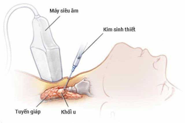
- Before surgery thyroidectomy for thyroid cancer, fine needle aspiration cytology - thyroid FNA; Long-term follow-up after thyroidectomy
3. Thyroid ultrasound technical procedure
The sonographer moves the transducer at a moderate speed, with continuous slices and scans in a cross-sectional plane, cutting along the thyroid axis to examine the entire lobes, isthmus and groups neighboring nodes.
When the doctor detects diffuse lesions, determine the length, width, thyroid thickness, and isthmus thickness, the acoustic structure compares with the adjacent muscle group and use color Doppler and Doppler energy assessment of angiogenesis.
When detecting lymph node involvement, it is necessary to determine the site of mapping, size, morphology and structure of lymph nodes
4. Benefits of Thyroid Ultrasound
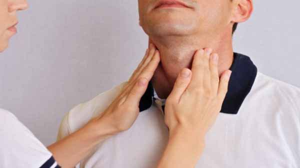
The thyroid gland is in a position that is easy to check and some diseases can be detected with the naked eye. Thyroid ultrasound should be examined during the general physical examination and indicated when the clinician examines the neck for suspicion of a neck tumor, a goiter (goiter), an intramediastinal mass, or when Clinical examination is normal but thyroid disease is suspected on laboratory tests.
Thyroid gland has many important functions and thyroid ultrasound is done easily and quickly, giving accurate results. Periodic examination and follow-up is recommended to detect pathological conditions and timely remedy, ensuring keep yourself and your family healthy.
To register for examination and treatment at Vinmec International General Hospital, you can contact Vinmec Health System nationwide, or register for an online examination HERE
MORE
Diagnosis of hypothyroidism by radiation Thyroid imaging Common thyroid diseases Role of thyroid scintigraphy in the diagnosis of thyroid diseases
Dịch vụ từ Vinmec
-
 Cách điều trị viêm tuyến giáp mãn tính
Viêm tuyến giáp mạn tính, hay còn gọi là viêm giáp Hashimoto, thường dẫn đến hậu quả suy giáp khi tuyến giáp bị tổn thương và mất dần khả năng sản xuất hormon. Vậy bệnh viêm tuyến giáp mãn tính ...
Đọc thêm -
 Viêm tuyến giáp mãn tính nên điều trị như thế nào?
Tôi đi khám ở bệnh viện và nhận được kết quả là bị đa nhân tuyến giáp, kích thước 0,4mm. Bác sĩ chỉ định tôi thử máu, sinh khiết và kết quả là viêm tuyến giáp mãn tính.
Đọc thêm -
 Nguyên nhân gây suy giáp ở người lớn
Suy tuyến giáp ở người trưởng thành là bệnh tương đối phổ biến, đặc biệt là phụ nữ. Suy tuyến giáp ở người lớn do nhiều nguyên nhân gây ra với biểu hiện lâm sàng thường thấy là bướu to ...
Đọc thêm -
 U tuyến giáp chuyển sang cường giáp có thể phẫu thuật không?
Cháu bị u tuyến giáp chuyển sang cường giáp. Vậy bác sĩ cho cháu hỏi u tuyến giáp chuyển sang cường giáp có thể phẫu thuật không? Phẫu thuật thì có thể điều trị dứt điểm không
Đọc thêm -
 Công dụng thuốc Betawtodex
Thuốc Betawtodex được bào chế dưới dạng viên nén, chứa thành phần chính là Betamethason. Thuốc được sử dụng trong điều trị một số căn bệnh có đáp ứng với corticoid.
Đọc thêm