
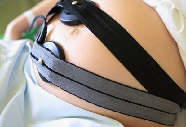
Monitor fetal health with CTG
What is CTG measurement? Measuring CTG is measuring fetal heart rate and uterine contractions with a fetal heart monitor called obstetric monitoring. The obstetrical monitor (or EFM) refers to the simultaneous recording of fetal heart rate and uterine contractions. The resulting plot is called a Cardiotocogram (CTG).
1. Normal and abnormal fetal heart curve
If from 120-160 beats/minute for term pregnancy: Normal; If >160 beats/min: Tachycardia; Basal fetal heart rate between 100-120 beats/min: Suspicious; Basal fetal heart rate < 100 beats/min: Bradycardia. What is abnormal fetal heart curve? If it's over 160 beats, it's called tachycardia, or if it's less than 100 beats, it's called deceleration. In the range of 100-120, there are suspicious signs:
Bradycardia: the cause of the bradycardia is due to the mother taking drugs (lowering blood pressure), the mother has low blood pressure or shock, convulsions, hypothermia, premature rupture of membranes, premature rupture of membranes, umbilical cord compression, placental abruption, preterm pregnancy, fetal arrhythmia, complete atrioventricular block; Tachycardia: Tachycardia is often related to the fetus's ability to cope with some threat to health. Tachycardia without increased rhythms with decrease or loss of intrinsic oscillation, or late deceleration indicates severe fetal hypoxia; Causes of fetal tachycardia include: maternal fever, anxiety, hyperthyroidism, chorioamnionitis, fetal anemia, fetal viral infection or infection, fetal hypoxia, after a prolonged deceleration, after external anesthesia dura, cardiovascular pathology;
Shift of the Fetal baseline: Shift of the Fetal baseline can take place in an upward or downward direction. If ascending, it may be due to an infection in the uterus, the fetus is hypoxic from any cause (umbilical cord compression). Shift in the basal fetal heart rate during the 2nd stage of labor is often associated with low cord blood pH; Basal line undulation: Severe fetal bradycardia may be seen in cases of umbilical cord circulation obstruction, placental abruption, or maternal complications such as hypotension, shock, convulsions, uterine rupture, or myocele supply is overstimulated. In cases where the undulating baseline is present within a limited period of the normal fetal heart rate, it may also reflect fetal neurological damage; Baseline unknown: The baseline fetal heart rate could not be determined. The reason for the unknown baseline could be a series of increased beats, intrinsic hyperoscillation, successively variable decelerations, or fetal arrhythmia. Increased rate: Is an indicator of a healthy fetus, also known as a responsive fetal heart rate curve;

2. Some tests to assess fetal health by CTG
If you have unusual symptoms, you should be examined and consulted with a specialist.
Để đặt lịch khám tại viện, Quý khách vui lòng bấm số HOTLINE hoặc đặt lịch trực tiếp TẠI ĐÂY. Tải và đặt lịch khám tự động trên ứng dụng MyVinmec để quản lý, theo dõi lịch và đặt hẹn mọi lúc mọi nơi ngay trên ứng dụng.
Dịch vụ từ Vinmec
-
 Thai nhi 34 tuần xuất hiện giãn bể lớn hố sau nên làm gì?
Chào bác sĩ. Cháu nhà tôi được 34 tuần, từ tuần thứ 30 khi đi siêu âm đã có kết quả là giãn đơn thuần bể lớn hố sau đường kính 12mm nhưng khi đó gia đình tôi không để ...
Đọc thêm -
 Sử dụng viên uống chống nắng có ảnh hưởng đến thai nhi?
Chào bác sĩ! Bác sĩ cho mình hỏi mình sử dụng viên uống chống nắng trong thời gian có thai thì liệu có ảnh hưởng gì đến thai nhi không? Mong bác sĩ tư vấn giúp mình. Mình xin cảm ...
Đọc thêm -
 Mẹ mắc bệnh Thalassemia cần làm gì sinh con được khỏe mạnh?
Chào bác sĩ. Em mắc bệnh Thalassemia, em có mang bầu bé thứ nhất đến tháng 6 em phát hiện bị dư ối tràn dịch màng phổi phải vào tim của thai nhi, em có đi khám và điều trị ...
Đọc thêm -
 Hỏi đáp: Mang thai có chụp được MRI ở não không?
Chào bác sĩ. Hiện mình đang mang thai được 27 tuần, mình mang thai có chụp được MRI ở não không? Chụp MRI não có ảnh hưởng gì đến thai nhi không? Cảm ơn bác sĩ đã tư vấn giúp ...
Đọc thêm -
 Đái tháo đường thai kỳ, mối bận tâm của mẹ bầu?
Đái tháo đường thai kỳ là một loại bệnh đái tháo đường phát hiện trong thai kỳ. Xảy ra khi cơ thể của mẹ bầu không thể sản xuất đủ insulin để đáp ứng nhu cầu của cả mẹ bầu ...
Đọc thêm