
The image of calcifications on ultrasound of the mammary gland
1. What is mammary calcification?
When it is not possible to give a specific etiology, the description of breast calcifications should include both their morphology and distribution. However, they should be reported if the physician reading the film is concerned that other film readers may misinterpret them.
2. What is breast ultrasound?

Mammography can be performed in the usual way by direct examination by a doctor or by 3D automatic breast ultrasound. With the 3D automatic breast ultrasound method, the patient will be examined by an automatic scanner. The specialist will then analyze the resulting images to look for abnormalities of the mammary glands.
The purpose of breast ultrasound is to detect lesions of the mammary glands. Breast ultrasound is being applied worldwide for diagnosis, early detection and monitoring of mammary gland abnormalities, especially breast cancer.
3. Benign breast tumor on ultrasound
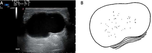
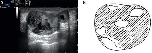
Clear, tender margins; Limitation of the lesion: The face is clearly and thinly separated from the adjacent healthy tissue (additional sign); Calcification: Coarse calcification; Direction: Parallel; Echoic structure: Drum (cystic) echo, thick echo, posterior tone change: hyperechoic, homonymous or slightly hypoechoic compared with mammary parenchyma; Has a thin shell; An ellipse in which the major axis is parallel to the skin surface; The block does not cause tension in neighboring structures; There are no signs suggestive of malignancy; Classification of benign breast tumors:
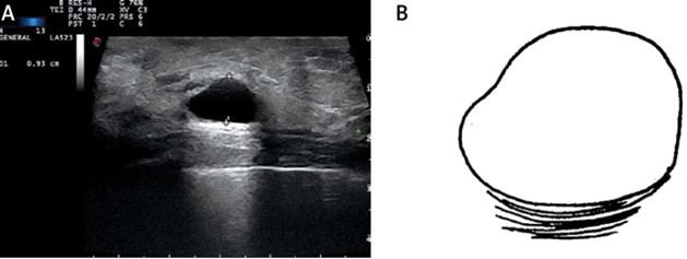
Type 1- Simple cyst: Round or oval cyst, clear margin, negative void, with increased illumination of the posterior wall;
4. Characteristics of malignant breast tumors
5. Where is a good breast ultrasound?

The early stage breast cancer screening package 2020 will help a lot in the treatment process. Early intervention by methods such as using drugs, radiation therapy, chemotherapy, surgery, hormonal therapy, biology... can minimize the effects of the disease, slow down the progression of the disease, especially saving the lives of breast cancer patients.
Currently, on the Vinmec International General Hospital system, there are modern breast ultrasound machines to help diagnose early calcified nodules. Vinmec uses 3D breast ultrasound device Invenia ABUS - the most advanced automatic ultrasound device in the world today in breast cancer screening with many outstanding advantages compared to other ultrasound techniques:
The system is capable of screening, diagnosing, monitoring before, after, treating breast diseases and detecting breast cancer with accuracy up to 90%. In addition, the mammography system helps diagnose early breast calcifications, increasing the ability to detect breast cancer at a very early stage when there are no clinical manifestations; Integrating ARFI tissue elastic technology, assessing tissue stiffness helps verify and complement the results on 3D mammograms, increasing accuracy in the diagnostic process; The 3D breast ultrasound system helps to create anatomical sections that are currently not possible with traditional ultrasound systems; Detect and identify small lesions to support effective disease monitoring and treatment; There are data to assess breast lesions quickly, safely because it is non-invasive and non-radioactive, so pregnant women can use this system.
Để đặt lịch khám tại viện, Quý khách vui lòng bấm số HOTLINE hoặc đặt lịch trực tiếp TẠI ĐÂY. Tải và đặt lịch khám tự động trên ứng dụng MyVinmec để quản lý, theo dõi lịch và đặt hẹn mọi lúc mọi nơi ngay trên ứng dụng.
Dịch vụ từ Vinmec
-
 Núm vú nổi cục sưng đỏ là dấu hiệu bệnh gì?
Tự dưng ở dưới núm vú em xuất hiện cục và sưng đỏ. Em sờ vào thấy cứng và hơi đau khi đụng vào. Vậy bác sĩ cho em hỏi núm vú nổi cục sưng đỏ là dấu hiệu bệnh ...
Đọc thêm -
 Kiểm tra u vú lành tính hay ác tính bằng kỹ thuật chẩn đoán nào?
Em là nam giới nhưng bị u vú 1 năm nay rồi, kích thước tầm 3cm không đau, không triệu chứng gì nên em cũng không đi khám. Bây giờ, em muốn khám xem u lành hay u ác. Vậy ...
Đọc thêm -
 Nhân tuyến vú kích thước 3.5cm có phải u vú không?
Tôi 50 tuổi, đi siêu âm vú có nhân 0.3 cm điểm 3 giờ, cách núm vú trái 1.5 cm, đã làm sinh thiết tế bào, không bị sao nhưng gần đây tôi thấy ở điểm 1 giờ cách núm ...
Đọc thêm -
 Đau vú trái, nặn núm vú ra dịch trắng là bị làm sao?
Năm nay cháu 21 tuổi. Tình trạng là 2 tuần nay cháu bị đau bên vú trái, ấn vào thì như bị sưng bên trong ạ. Cháu có nặn núm vú thì tiết ra nước trắng trong còn bình thường ...
Đọc thêm -
 Nổi cục ở núm vú có phải ung thư vú không?
Em có sờ thấy 1 cục nhỏ bằng hạt lạc không đau, không di chuyển ở gần núm vú. Vậy bác sĩ cho em hỏi nổi cục ở núm vú có phải ung thư vú không? Em cảm ơn.
Đọc thêm