
What is a straight chest X-ray?
1. Straight chest X-ray
X-ray is the most important laboratory test in the diagnosis of lung disease. The value of radiographs in the diagnosis of lung disease is second only to the diagnosis of bone disease. A chest X-ray will give the clearest picture of the structure of the heart, lungs or blood vessels, joints, bones, and spine behind the chest.
A straight chest radiograph can identify underlying lung damage and heart-related problems. From here, the doctor will determine the relationship of that lesion to the surrounding organs: mediastinum, ribs and intercostal space, trachea, diaphragm,... The meticulous analysis of the relationship This will avoid confusion in the diagnosis of cardiopulmonary diseases. X-ray film will see the fluid in the lungs, or the empty space around the lungs, the heart structure, the basis for determining pneumonia, or cancer...
Cardiopulmonary x-ray is one of the things. The first method doctors think of when a patient has suspicious signs such as: chest pain, fever, persistent cough, shortness of breath, ... are assigned to take X-rays for diagnosis.
2. What are the effects of chest X-ray?
Can accurately determine the location of damage to the lobe or segment, or signs of suspected lung disease, in the thorax,... Monitor the progress of health status, level damage to the lungs, heart, or chest cavity. Estimating the rate of cardiac tamponade or invasion to the esophageal margin Identify the lesion in the mediastinum or suspect the presence of a tumor. Detecting unusual symptoms in the lungs, in the heart. Monitor activity in the heart and lungs over time during medical treatment. Straight chest X-ray is considered a popular diagnostic aid at the moment, aiming to detect early abnormalities in the heart, lungs and neighboring functional areas.
The principle of a straight chest X-ray is to use X-ray radiographs to evaluate imaging results in both functional organs and body tissues. Organs can absorb or block X-rays differently, but their nature is different, creating areas of light and dark, dark and light; This has the positive side of making it easier for the physician to delineate the area or structure more clearly.
3. When do you need a straight chest X-ray?

When the patient has signs of disease related to cardiopulmonary function, the diagnosis will need to be made based on a straight chest X-ray film. If the results show that there are signs of disease development, the patient needs to be assigned a specific regimen and start treatment early, so that the disease will last for a long time and will encounter unpredictable complications later.
Normally, a straight chest X-ray is examined and clearly indicated by a doctor, only when it is suspected that the patient has an abnormality in heart or lung activity.
Subjects that need a straight chest X-ray are people who:
Chest trauma with a risk of rib fractures Pulmonary contusion Lung cancer Pneumonia Chronic obstructive pulmonary disease Pneumothorax Cardiovascular disease
4. The role of straight chest X-ray
This method will help the doctor see more accurately visual images of internal organs that cannot be seen with the naked eye, thereby being able to diagnose and advise properly according to the patient's health status.
Based on the X-ray film, the doctor can see the change:
Detecting signs of abnormality in the lungs: X-rays will help identify lung infections or cancer. Cardiac abnormalities related to the lungs: Fluid can be seen to accumulate in the lungs causing pulmonary edema, which is caused by abnormalities of the heart. Heart size: the shape and size of the heart, if abnormal, are signs of an impending underlying disease, such as: heart failure, congenital heart disease, pericardial effusion, regurgitation heart valve or other problems with the vessels and heart valves... Transport of blood vessels: Through the film, the large vessels around the heart can be clearly seen on X-ray films such as: arteries, aorta, veins.. ., suspected when there is an enlarged artery in the artery or another problem related to blood flow... Calcification: Calcium in the heart or in the blood vessels when present will cause adverse effects on the heart and lungs. . Cardiac calcification can affect the functioning of the heart valves, the aorta, the myocardium or the protective membrane of the heart. Calcification of the lungs, usually from an untreated infection or another serious lung problem. After CPR: An X-ray after a CPR or other chest surgery can diagnose a cardiopulmonary abnormality that has occurred. Because after open surgery, the internal organs are exposed to the air environment, so it is easy to encounter infection or complications if the incision is not carefully aseptic. Monitoring the body's recovery after open heart, lung or esophageal surgery...
5. Conduct a straight chest X-ray
First, the X-rays will in turn penetrate the body's structures in different directions. When taking a straight chest X-ray, the image on the film will show: Long bones are dense and dense, so they appear bright white, the heart has the ability to block some radiation, so it will have a lighter color, The lungs contain air, so they form a small dark mass.
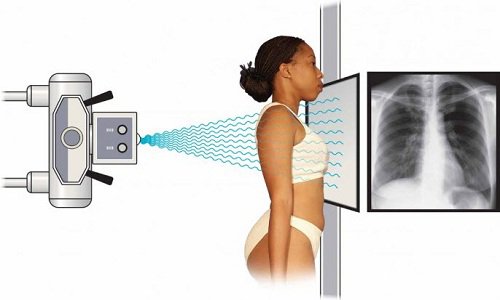
Move into the imaging room under the guidance of the technician The technician will guide you to stand next to a plate containing X-ray film inside or a special receiver that can record images Enter the computer The technician will teach you how to stand to take the picture. Hold your breath while taking to increase the sharpness of the X-ray image
6. Notes when taking a straight chest X-ray
You will carry out the doctor's orders: change, take off your shirt and put on a patient shirt already in it. Your doctor may ask you to remove jewelry, dental appliances, glasses, or metal objects that can interfere with the X-ray.
For female patients, you should tell your doctor and radiologist if they suspect you are or have become pregnant. Chest X-ray technique will not be performed during pregnancy to avoid radiation exposure to the fetus. But if your medical condition requires an X-ray, your doctor will take protective measures to reduce the risk of radiation exposure to your unborn baby.
Through the image of a straight chest X-ray film, the doctor will understand the patient's health status, give accurate advice or give a specific treatment plan and treat the patient early when there is a problem. Dangerous problems may occur.
Để đặt lịch khám tại viện, Quý khách vui lòng bấm số HOTLINE hoặc đặt lịch trực tiếp TẠI ĐÂY. Tải và đặt lịch khám tự động trên ứng dụng MyVinmec để quản lý, theo dõi lịch và đặt hẹn mọi lúc mọi nơi ngay trên ứng dụng.
Dịch vụ từ Vinmec
-
 Nguyên nhân và các yếu tố nguy cơ gây thuyên tắc mạch phổi
Thuyên tắc mạch phổi là căn bệnh thường gặp với tỷ lệ tử vong khoảng 30% nếu không được phát hiện và điều trị sớm. Có nhiều nguyên nhân gây bệnh, trong đó nguyên nhân thường gặp nhất là huyết ...
Đọc thêm -
 Sử dụng lưu lượng đỉnh kế chẩn đoán bệnh hen
Lưu lượng thở ra đỉnh là nghiệm pháp giúp đo lường tốc độ thở ra tối đa của một người, nhằm kiểm tra chức năng phổi và thường được sử dụng trong chẩn đoán bệnh hen.
Đọc thêm -
 Chẩn đoán và điều trị kịp thời thuyên tắc mạch phổi
Thuyên tắc phổi là tình trạng một cục máu đông lọt vào mạch máu trong phổi, ngăn chặn dòng lưu thông bình thường của máu trong vùng đó, gây trở ngại cho việc trao đổi không khí và có thể ...
Đọc thêm -
 Liệu pháp oxy lưu lượng cao qua ống thông mũi (High Flow Nasal Cannula) - Phần 3
Liệu pháp oxy lưu lượng cao qua ống thông mũi có thể làm giảm đáng kể cả tỷ lệ tử vong và tỷ lệ đặt nội khí quản ở bệnh nhân suy hô hấp cấp có suy giảm miễn dịch, ...
Đọc thêm -
 COVID-19 ảnh hưởng thế nào đến người mắc bệnh phổi tắc nghẽn mãn tính (COPD)?
Virus SARS-CoV-2 gây ra bệnh COVID-19, có thể dẫn đến các vấn đề hô hấp từ nhẹ đến nghiêm trọng. Do có sẵn bệnh lý nền về phổi, người mắc Covid khi bị COPD nhiều nguy cơ diễn tiến nặng ...
Đọc thêm