
Diagnostic imaging of neuroinfection
1. What is a nerve infection?
The most prominent symptoms commonly seen in patients with CNS infections are fever, headache, sensory disturbances, Kernig and Brudzinski signs, neck stiffness, and abnormalities in the CSF. . In general, all of these symptoms are very rarely present at the same time in a particular disease. Just the presence of one of the typical symptoms above can suggest that the patient is suffering from a neurological infection.
Neuroinfection is evaluated as a medical emergency, and the patient should be taken to the right diagnostic steps to pinpoint the specific cause of the disease. Usually, the diagnostic procedures for neuroinfection will include: Take the history - perform a physical examination - order the patient to perform a complete blood count test - blood culture - lumbar puncture and do laboratory tests. for cerebrospinal fluid culture, chest X-ray.
For cerebrospinal fluid, the specialist will count the number of cells as well as sugars and proteins and stain the slide for bacteria (including acid-resistant bacteria if necessary). , cultures for pyogenic bacteria and for tuberculosis and fungi if indicated.

2. Diagnostic imaging of neuroinfection
2.1 Herpes encephalitis
Herpes encephalitis has a relatively high mortality rate, so early diagnosis plays a very important role in saving patients' lives.
Use diagnostic imaging to reflect the lesional nature of the disease. In which, CT is rarely used because of its low sensitivity, it can show the hypodense images of the bilateral temporal lobes.
On the MRI image of the lesion, there is hypointensity on T1W, hyperintensity on T2W, and hyperintensity on FLAIR appearing in the first 48 hours. In particular, the diffusion pulse sequence (DWI/ADC) is considered to be the most sensitive to images of limited cell diffusion due to edema and toxicity. The DWI index is closely related to disease activity and response to treatment. On MRI images with injection of magnetic contrast, increased enhancement of the lesion is usually not found in the early stages and often gyrus enhancement is detected in later stages.

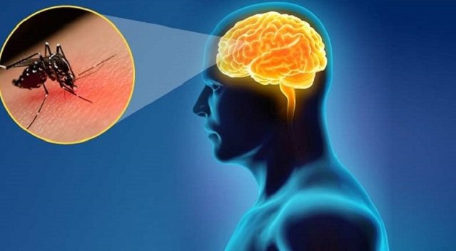
2.2 Japanese Encephalitis
Imaging diagnosis of Japanese encephalitis on CT images has symmetrical or asymmetrical hypodense images of bilateral hippocampus. On MRI, brain lesions are often hypointense on T1W, hyperintensity on T2W/Flair, and diffusely limited with hyperintensity on DWI.
Japanese encephalitis can include lesions in the cerebellum, cerebral cortex as well as the basal ganglia. In addition, bleeding often occurs. In general, bilateral hippocampal hemorrhage is quite specific in Japanese encephalitis.
2.3 HIV encephalopathy
HIV encephalopathy without enhancement or space-occupying effect. On CT images, white matter lesions can be seen with reduced attenuation when many abnormalities are present.
In addition, studies showed diffuse diffuse symmetric white matter bilaterally on FLAIR and not subcortical white matter.
It is necessary to distinguish precisely from other white matter-related diseases such as:
● White matter lesions in small vessel disease.
White matter disease caused by intoxication .
White matter encephalopathy due to anemia or lack of oxygen .
2.4 CYTOMEGALOvirus
CMV imaging shows diffuse white matter abnormalities, cortical atrophy, or hyperintensity on FLAIR or periventricular hyperintensity.
2.5 CMV newborn
Definitive imaging in today's advanced imaging methods helps to evaluate calcification, abnormal neuronal migration, or periventricular cysts, hydrocephalus or corpus callococcal dysplasia, cortical hypoplasia .
In general, nerve infection comes from many different causes, can cause many dangerous complications if not diagnosed and treated promptly. Today, with the development of modern medicine, imaging methods have contributed to accurately assessing the condition of the lesion as well as the stage of disease development, contributing to the appropriate treatment regimen. .
However, neuroinfection imaging requires advanced medical equipment along with a team of highly qualified and experienced doctors. Therefore, to receive the best diagnosis and treatment, you should choose the top medical centers in our country. Currently, the Department of Diagnostic Imaging is one of the spearheads of Vinmec International Hospital, which is equipped with the most modern and advanced equipment in the country as well as in the region. The department has 6 ultrasound rooms, 4 DR X-ray rooms (1 full-axis machine, 1 brightener, 1 synthesizer and 1 mammography machine), 2 DR mobile X-ray machines, 2 cutting rooms. multi-row computer class with receiver (1 128-series and 1 16-series), 2 Magnetic Resonance imaging rooms (1 3 Tesla machine and 1 1.5 Tesla machine), 1 interventional angiography room with 2 planes and 1 room measure bone mineral density. The doctors at the Faculty are well trained at home and abroad. Especially when coming to Vinmec International General Hospital, patients do not need to wait long, have a quick examination time, and are carefully guided by a team of experienced medical staff on the use of medicines and instructions. Lead positive lifestyle changes so that patients can easily adhere to their treatment management.
Master Doctor Le Anh Viet has 07 years of experience working in the field of diagnostic imaging, performing well in diagnostic imaging techniques. Currently, the doctor is working at the Department of Diagnostic Imaging and Nuclear Medicine - Vinmec Times City International General Hospital.
If you still have any unanswered questions related to neuroinfection imaging, you can directly contact Vinmec International Hospital system nationwide for specific advice. .
Để đặt lịch khám tại viện, Quý khách vui lòng bấm số HOTLINE hoặc đặt lịch trực tiếp TẠI ĐÂY. Tải và đặt lịch khám tự động trên ứng dụng MyVinmec để quản lý, theo dõi lịch và đặt hẹn mọi lúc mọi nơi ngay trên ứng dụng.
Dịch vụ từ Vinmec
-
 Trẻ 11 tháng tuổi sau bó bột gãy xương đùi, mối xương chồng lên nhau nhau có sao không?
Bé trai nhà cháu 11 tháng tuổi bị gãy xương đùi trái hiện đã được bó bột. Chụp X quang sau khi bó bột hình ảnh chính diện xương thẳng, hai mối xương chồng lên nhau. Hình ảnh từ phải ...
Đọc thêm -
 Sốt nhẹ sau khi chụp X quang đường mật có thuốc cản quang có sao không?
Tôi mới chụp X quang đường mật có thuốc cản quang thì bị ớn lạnh sốt nhẹ. Xin hỏi bác sĩ sốt nhẹ sau khi chụp X quang đường mật có thuốc cản quang có sao không? Cảm ơn bác ...
Đọc thêm -
 Chụp X quang nhiều lần có bị nhiễm tia X không?
Vừa qua, em đi khám bệnh xương khớp, bác sĩ chỉ định em chụp X quang vai trái phải thẳng nghiêng, cột sống cổ, cột sống thắt lưng, chụp phổi thẳng nghiêng,.... Khi chụp xong em hoảng hốt khi thấy ...
Đọc thêm -
 Chụp x quang có ảnh hưởng đến thai nhi không?
Chào bác sĩ! Em vừa mới có bầu 6 tuần nhưng em nhớ ra khoảng 3 tuần trước em có chụp X quang. Bác sĩ cho em hỏi như thế có ảnh hưởng đến thai nhi không ạ? Mong bác ...
Đọc thêm -
 Chụp X quang vú có chẩn đoán được ung thư vú không?
Bác sĩ cho em hỏi chụp X quang vú có chẩn đoán được ung thư vú không? Độ chính xác bao nhiêu % ạ? Thời gian chụp ở Vinmec có lâu không? Cảm ơn bác sĩ tư vấn.
Đọc thêm