Risk stratification and prognostic prediction of GISTs by data visualization techniques
Datagrammetry is based on mathematical quantification of image heterogeneity, through analysis of the distribution and relationship of pixel intensities in a region of interest (ROI). Analysis of data-forming images requires a multi-step process, starting with image acquisition, and including lesion segmentation, feature extraction, feature selection and reduction, building predictive models, and ultimately validate and interpret clinical results.
1. Risk stratification and prognostic prediction of GISTs by data visualization techniques
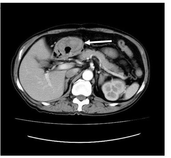
2. Datagram imaging models have demonstrated that improved preoperative prediction of high-risk GIST compared with conventional imaging assessment
3. The potential of data visualization
Prediction of malignancy potential or postoperative recurrence-free survival In two subsequent studies, Chen et al built a support vector machine and a residual neural network-based model to predict potential malignancy or recurrence-free survival 3 years and 5 years after focal total resection of GIST, respectively. In those studies, the Authors enrolled a cohort of medical patients for model training, which was then validated in internal and external groups, with performance ranging from good to close. as perfect for GIST risk stratification and prediction of recurrence-free survival at 3 years and 5 years, respectively, Structural feature survival analysis of disease progression was also performed. by Ekert et al. on contrast-enhanced CT, whereas only one study performed data visualization on MRI. Fu et al extracted texture features from T2, DWI and ADC weighted map images to determine the prognosis of metastatic GIST, reporting that texture features on DWI and ADC maps have correlated well with overall survival.
Finally, the Ki67 index represents a marker of tumor cell proliferation, which is also associated with poor prognosis in GISTs. In a study of 339 GISTs, the Contrast-enhanced CT angiography marker demonstrated a significant correlation with Ki67 expression, providing an additional value for prognostic assessment.
Để đặt lịch khám tại viện, Quý khách vui lòng bấm số HOTLINE hoặc đặt lịch trực tiếp TẠI ĐÂY. Tải và đặt lịch khám tự động trên ứng dụng MyVinmec để quản lý, theo dõi lịch và đặt hẹn mọi lúc mọi nơi ngay trên ứng dụng.
References Cannella R, La Grutta L, Midiri M, Bartolotta TV. New advances in scintigraphy of gastrointestinal stromal tumors. World J Gastroenterol 2020; 26 (32): 4729-4738 [PMID: 32921953 DOI: 10.3748 / wjg.v26.i32.4729 ]
Dịch vụ từ Vinmec
-
 Chụp cắt lớp vi tính ở người mắc bệnh cơ tim
Bệnh cơ tim là nguyên nhân chủ yếu dẫn đến suy tim, có tiên lượng xấu và tỷ lệ tử vong khá cao. Chẩn đoán sớm có ý nghĩa quan trọng trong việc phát hiện và điều trị bệnh. Trong ...
Đọc thêm -
 Vai trò của siêu âm, chụp cắt lớp vi tính trong chẩn đoán, điều trị bệnh lý gan mật
Ngày nay con người đang phải đối mặt với nhiều vấn đề như thực phẩm nhiễm hóa chất, lạm dụng rượu bia, môi trường ô nhiễm,... khiến tỷ lệ bệnh nhân mắc các bệnh lý về gan mật ngày một ...
Đọc thêm -
 Quy trình chụp cắt lớp vi tính cột sống cổ có dựng hình 3D
Chụp CLVT cột sống cổ có dựng hình 3D là kỹ thuật chẩn đoán hình ảnh được áp dụng rộng rãi để giúp bác sĩ đánh giá được tình trạng thực tế của cột sống cổ, các bệnh lý đi ...
Đọc thêm -
 Làm thế nào để phát hiện sỏi thận chính xác nhất?
Bác sĩ cho em hỏi làm thế nào để phát hiện sỏi thận chính xác nhất? Em nên dùng siêu âm hay X quang thưa bác sĩ? Trong trường hợp em sử dụng PP siêu âm phát hiện ra sỏi ...
Đọc thêm -
 Những tiến bộ mới trong tạo hình dữ liệu hoá của các khối u mô đệm đường tiêu hóa
Mặc dù tạo hình dữ liệu hoá có tiềm năng nghiên cứu to lớn để cải thiện việc đánh giá định lượng khối u mô đệm đường tiêu hóa, nhưng có một số hạn chế thách thức việc ứng dụng ...
Đọc thêm