
Background erasing digital angiography with iodine contrast agent
1. What is an iodinated contrast digital pulmonary angiogram?
According to the Seldinger method, the entrance is from the femoral vein, possibly from the subclavian and internal jugular veins into the superior vena cava into the right atrium to the right ventricle and into the pulmonary artery. From there, it is possible to intervene in the patient's pathology such as: Embolism, thrombectomy, fibrinolysis...
2. When is digitized pulmonary angiography with iodine contrast indicated?
Indications for this pulmonary angiogram, although there is no absolute contraindication, but in some cases, the patient is in a state of severe organ failure (liver, kidney..), thrombosis in the right atrium, drug allergy to the patient. ophthalmology, pregnant women, doctors still have to carefully consider and evaluate each person's condition to decide whether to perform or not.

3. Background erasing digital pulmonary angiography with iodine contrast agent
3.1 Executor
Usually children under 5 years old or those who are not awake and do not cooperate with the doctor will do the anesthesia.
3.2 Asking the patient
● Need to fast before 6 hours. Can drink no more than 50ml of water.
3.3 Implementation process
● Anesthesia method
● Place the patient supine on the scanning table, place an intravenous line, pre-anesthesia or anesthetic if necessary (patient is not alert, young child...)
● Select technique and route Inlet of the catheter: According to the Seldinger method, the entrance can be from the femoral vein, brachial vein, and jugular vein. Usually the femoral vein, unless this route does not work, then choose other routes.

3.4 Performing the trick
● Anesthetize the site where the catheter is inserted into the lumen.
● Needle puncture, put the kit into the femoral vein.
● Insert the lead and angiogram through the catheter into the inferior vena cava, up the right atrium, into the right ventricle, and up the aorta.
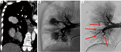
● Pulmonary angiography with contrast agent through a syringe can show the entire right and left pulmonary artery system, identify abnormalities: Aneurysm, malformation, thrombosis...
● Obstruction, abnormal recanalization usually by means of embolization, angioplasty, and specialized thrombectomy.
● Withdraw the catheter and tube into the lumen after satisfactory angiography and intervention, apply gentle pressure by hand to stop bleeding for 15 minutes, then apply pressure for 6-8 hours.
● Select the catheter technique and access route: According to the Seldinger method, the entrance can be from the femoral vein, brachial vein, and jugular vein. Usually the femoral vein, unless this route does not work, then choose other routes.
● The nurse disinfects the groin area on both sides (the way to the femoral vein). Stretch, try to cover the entire patient, leave open the puncture site, the place where the catheter is inserted into the angiogram.
● Anesthetize the site where the catheter is inserted into the lumen.
● Needle puncture, put the kit into the femoral vein.
● Insert the lead and angiogram through the catheter into the inferior vena cava, up the right atrium, into the right ventricle, and up the aorta.
● Pulmonary angiography with contrast agent through a syringe can show the entire right and left pulmonary artery system, identify abnormalities: Aneurysm, malformation, thrombosis...
● Obstruction, abnormal recanalization usually by means of embolization, angioplasty, and specialized thrombectomy.
● Withdraw the catheter and tube into the lumen after satisfactory angiography and intervention, apply gentle pressure by hand to stop bleeding for 15 minutes, then apply pressure for 6 hours.
● After finishing the digital pulmonary angiography procedure with iodine contrast, the patient lies on the bed to rest, the nurse will monitor the dorsal pulse, monitor bleeding, hematoma at the puncture site and Monitor the whole body (pulse, blood pressure, patient response).
Vinmec International General Hospital has applied the technique of digital pulmonary angiography to erase the background in the examination, diagnosis and detection of diseases for patients. All medical examination and examination procedures are performed by a team of doctors with many years of experience. Combined with that is a system of modern machinery and equipment. Thanks to good medical conditions, patients can be assured of the diagnosis results as well as the treatment direction when prescribed by the doctor.
Master. Dr. Le Hong Chien has many years of experience working in the field of diagnostic imaging and interventional radiology (endovascular intervention and extravascular intervention). Before being a Doctor of Diagnostic Imaging - Intervention at Vinmec Times City International Hospital, Dr. Chien was a Doctor at the Department of Diagnostic Imaging, Hospital 19.8 - Ministry of Public Security and Hong Ngoc General Hospital. .
Any questions that need to be answered by a specialist doctor as well as customers wishing to be examined and treated at Vinmec International General Hospital, you can contact Vinmec Health System nationwide or register online HERE.
What is pulmonary hypertension? How dangerous is pulmonary hypertension? Overview of common diseases in the lungs
Dịch vụ từ Vinmec
-
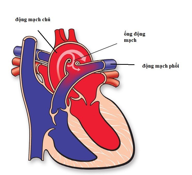 Điều trị ống động mạch hình phễu chưa tăng áp phổi ở trẻ như thế nào?
Chào bác sĩ! Bé nhà em năm nay 31 tháng tuổi nhưng cháu còn ống động mạch dạng hình phễu kích thước của ống ở phía của động mạch chủ xuống 5.6mm, phía động mạch phổi 3mm, chiều dài 6,2 ...
Đọc thêm -
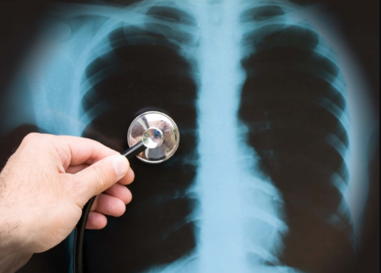 Nên làm gì khi phát hiện động mạch phổi to?
Em mới đi chụp Xquang ngực kiểm tra phổi, thấy động mạch phổi to. Mong bác sĩ tư vấn giúp tôi về tình trạng này. Tôi xin cảm ơn.
Đọc thêm -
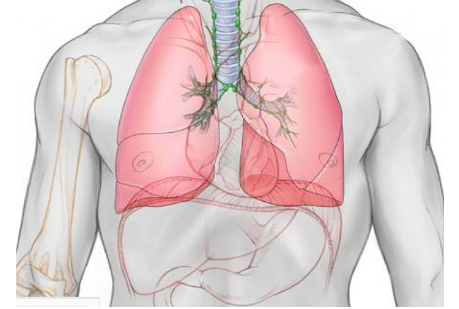 Chụp số hóa xóa nền động mạch phổi
Động mạch phổi là mạch máu có chức năng dẫn truyền máu đến phổi và từ phổi đi các cơ quan khác. Chụp số hóa xóa nền động mạch phổi sẽ giúp bác sĩ thu thập hình ảnh về bộ ...
Đọc thêm -
 Có cần điều trị hở van tim 1/4 và bệnh nguy hiểm không?
Chào bác sĩ! Vợ tôi đi siêu âm tim ở bệnh viện thấy có kết quả hở van tim 1/4. Bác sĩ cho tôi hỏi, có cần điều trị hở van tim 1/4 và bệnh nguy hiểm không? Tôi xin ...
Đọc thêm -
 Vinmec có chụp động mạch phổi không?
Chào bác sĩ, em muốn hỏi ở Vinmec có chụp động mạch phổi không? Có kỹ thuật nào nhanh có kết quả mà chính xác không ạ?
Đọc thêm