
Current indications for mammography magnetic resonance imaging
Magnetic resonance mammary gland plays an increasingly important role with high sensitivity, so it is valuable for early diagnosis of breast cancer. From there, it can be detected at an early stage of the disease and the patient can be completely cured.
1. Purpose of a mammogram
In recent years, breast disease is more and more common and more concerned by the community. Besides the two basic diagnostic imaging methods, namely breast ultrasound and mammogram, which are valuable in screening for breast cancer, Magnetic Resonance is a technique that is increasingly used in the field of disease diagnosis. Breast management, it has a role to solve the cases of unclear detection by the above two methods and clinical difficulties such as hidden breast cancer.
Breast cancer is on the rise and is the leading cause of death in women. Currently, there is no vaccine to prevent breast cancer, so the best method to protect yourself is to periodically self-screen for breast cancer at specialists and mammography centers for early detection. In the early stages and with the right treatment, the disease can be completely cured.

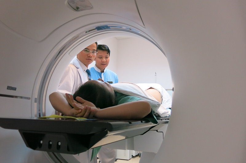
Mammography MRI will use a magnetic field to take many pictures of breast tissue, these images will be processed through a computer system to produce detailed, high-resolution images of the breast, helping doctors The doctor has the basis of analysis and makes an accurate diagnosis. Breast MRI has a high sensitivity of nearly 100% for the detection of invasive breast cancers of a few mm in size, with specificity ranging from 30-90%.
Breast MRI is indicated in the early detection of breast cancer in particular and breast diseases in general.
2. Indications for mammography
Determine the nature of lesions in inconclusive cases by means of routine mammography. Evaluation of the extent of the lesion in both adenocarcinoma and invasive lobular carcinoma. Invasive assessment of the muscle fascia. Examination of the contralateral breast in patients with breast cancer. Evaluation before, during and after the treatment, Evaluation of residual damage after tumor removal surgery.
Suspected tumor recurrence in patients with or without postoperative mammography. There is malignant axillary lymph node involvement with unknown primary tumor. Breast cancer screening in high-risk patients (especially in patients with suspected or proven BRCA gene mutations). Examine very large breasts with or without breast implants.
3. Notes for accurate results when examining breast MRI
Facilities: Magnetic resonance machine with high force (currently Vinmec Hai Phong General Hospital has a 3.0 Tesla machine) and specialized breast surface coil. Patient preparation: For patients, screening for breast disease is usually done in the first week after menstruation. Patients with suspected cancer can do it at any time. Tell your doctor what medications you are taking and if you have a history of any allergies. Are you pregnant? Do you have metal objects on you? The patient must be explained the scan procedure to avoid surprises and cooperate well with the doctor. People: Doctors and technicians must be well-trained to avoid performing the wrong technique for substandard imaging results and inaccurate lesion identification.
Technique: make successive cuts of less than 3mm thickness with a resolution of less than 1mm. Performing fat-free T1W pulses; T2W of non-injectable fat removal in the horizontal and vertical planes and sequence of T1W of pre-injection fat removal. Then perform injection of magnetic contrast agent (Gado) at 0.1-0.2 mmol/kg, then take T1W to remove fat after injection and take 2 to 5 additional pulse sequences after injection. These images are then processed to generate drug uptake curves, background removal images and post-injection peaks. All these images will be read and concluded by the radiologists.
Vinmec International General Hospital is one of the hospitals that not only ensures professional quality with a team of leading medical doctors, modern equipment and technology, but also stands out for its examination and consultation services. comprehensive and professional medical consultation and treatment; civilized, polite, safe and sterile medical examination and treatment space.
Customers can directly go to Vinmec Health system nationwide to visit or contact the hotline here for support.
Dịch vụ từ Vinmec
-
 Các phương pháp nội khoa trong điều trị ung thư vú
Ung thư vú là sự phát triển bất thường của các tế bào trong vú. Những tế bào này tăng trưởng và phát triển thành một khối ung thư có khả năng xâm lấn sang các bộ phận khác của ...
Đọc thêm -
 Các phương pháp can thiệp vú
Ở Vinmec hiện tại có 2 chương trình: Can thiệp để chẩn đoán và can thiệp vú để điều trị: Can thiệp vú chẩn đoán gồm có: Chọc tế bào, sinh thiết kim lõi, sinh thiết hút chân không và ...
Đọc thêm -
 Chuẩn bị trước khi chụp MRI vú
Chụp cộng hưởng từ vú hoặc chụp MRI vú là kỹ thuật hình ảnh được sử dụng để phát hiện ung thư vú và các bất thường khác ở vú. MRI vú thường được thực hiện sau khi bạn sinh ...
Đọc thêm -
 Điều gì làm nên sự khác biệt của trung tâm ung bướu xuất sắc (COE) và lợi ích dành cho người bệnh Vinmec
Hiện nay, Hệ thống Y tế Vinmec đang triển khai xây dựng Trung tâm Ung bướu xuất sắc (COE) đầu tiên ở Việt Nam với sự hợp tác của các chuyên gia tại Đại học Pennsylvania (Top 8 Đại học ...
Đọc thêm -
 Vì sao nên đi chụp x-quang vú để sàng lọc ung thư vú?
Chụp x-quang vú rất cần thiết vì có thể chẩn đoán sớm ung thư vú, từ đó sẽ giúp việc điều trị được tốt hơn, kéo dài thời gian sống cho bệnh nhân. Bên cạnh đó, khi đi chụp x-quang ...
Đọc thêm