
Diagnosis of Barrett's esophagus and esophageal cancer by artificial intelligence
1. Overview of artificial intelligence
In colonoscopy, AI has begun to aid in improving colon polyp detection and adenoma detection (ADR) rates, to distinguish between benign and precancerous lesions based on the interpretation of patterns their surface.
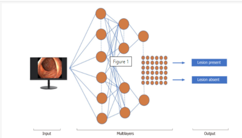
Machine learning (ML) and deep learning (DL) can be considered as subfields of artificial intelligence. ML is a form of artificial intelligence that can support decision-making, allowing improvements to applied algorithms without programming, including examining data and implementing descriptive models and models. predicted (Figure 1).
2. What is Barrett's esophagus?
3. The role of endoscopic diagnosis of Barrett's esophagus
It is important to document neoplastic changes in patients with Barrett's esophagus, and changes in endoscopic images have helped in the early detection of minimal neoplastic lesions based on individual features. mucosal differentiation.

4. Artificial Intelligence, Barrett's Esophagus and Esophageal Cancer
The endoscopic images were independently evaluated by five experts and then compared with the AI-powered probability map, showing strong correspondence. As the importance of manual segments differed significantly, their intersection was considered as the cancer region (C1 region) in each C1 image.

5. Some studies of AI in esophageal cancer diagnosis
In these early studies, the authors developed a CAD computer model and displayed promising performance scores in the classification/segmentation domains during the assessment of Barrett's esophagus.
However, these results are achieved using high-quality endoscopic images that are not always obtained during daily clinical practice. This system was previously developed to further speed up image analysis for classification and resolution of dense predictions, displaying the color-coded spatial distribution of cancer probabilities.
6. The role of Complex Neural Networks CNN in artificial intelligence

7. The role of the HD-WLE system and the NBI . system
Both the HD-WLE system and the NBI system detected 104/113 (92%) patients with intestinal metaplasia, but the NBI required fewer biopsies per patient and showed a higher detection rate of dysplasia significantly (30% vs. 21%). During endoscopic examination with NBI, all areas of high-grade dysplasia and cancer showed mucosal or vascular irregularities. Surface samples observed with the conventional NBI system did not contain high-grade dysplastic or cancerous lesions, suggesting that biopsy could be avoided in the following cases.
8. Using endoscopic trimodal imaging (ETMI) to detect early cancer in Barrett's esophagus
Van der Sommen et al used a computer algorithm to detect early cancerous lesions in Barrett's esophagus and used specific textures, color filters and based machine learning algorithms based on 100 images Photos from 44 patients with Barrett's esophagus. This system identifies early cancer lesions at the patient level with sensitivity and specificity of 86% and 87%, respectively. The author assumes that the automated computer algorithm implemented for this study can identify early cancerous lesions with reasonable accuracy.
De Groof et al developed a CAD system using endoscopic images of Barrett cancer based on endoscopic images of 40 Barrett cancerous lesions and 20 Barrett's esophagus without dysplasia, achieving sensitivity and The specificity for detecting such lesions is 95% and 85%, respectively.
9. Volumetric laser endoscopy (VLE) technology in esophageal cancer diagnosis
VLE can enhance detection of neoplastic lesions in Barrett's esophagus by performing circumferential scanning of the esophageal wall layers. Sixteen patients with Barrett's esophagus were included in the study and a total of 222 laser markers (LMs) were placed, 97% of them visible on WLE. All LMs were clearly visible on the VLE immediately after marking, and 86% were confirmed during post-school analysis. Laser targeting LM has 85% accuracy of traces. This initial study applied to humans suggests that VLE-guided LM may be a viable and safe procedure.
In another study, the same author used a database of VLE images from Barrett's esophagus with/without tumor, accurately correlating them with histology to develop scores VLE prediction. The receiving performance curve of this predictor shows an area under the curve (AUC) of 0.81. A value of ≥ 8 correlates with a sensitivity of 83% and a specificity of 71%.
10. The role of optical tomography (OCT) in the diagnosis of esophageal cancer
Evans et al., examined 177 OCT images from patients with the histological diagnosis of Barrett's esophagus. Histopathological analysis was IMC/HGD in 49 cases, LGD in 15, IGD in 8, specific IM in 100, while gastric mucosa in 5 patients. A significant correlation was found between the histopathology of mucosal cancer or high-grade dysplasia and the scores for each of the imaging features, extent of surface abnormalities, and glandular architecture. When using a dysplasia index ≥ 2 determination, 83% sensitivity and 75% specificity were determined for the diagnosis of IMC/HGD.
In a community health care center, 27 Barrett's esophagus patients who underwent 50 EMR mucosalectomy procedures were imaged with VLE and pCLE, and classified as cancerous/non-cancerous in the body histological results. The sensitivity and specificity of pCLE for the detection of Barrett's esophagus were 76% and 79%, respectively. OCT-SI showed a sensitivity of 70% and specificity of 60%. Furthermore, the VLE-DA technique showed a sensitivity of 86%, a specificity of 88% and a diagnostic accuracy of 87%.
Esophageal squamous cell carcinoma (SCC) is the sixth leading cause of death worldwide and a greater percentage affects developing countries due to delayed diagnosis. Esophageal endoscopy using Lugol dye is currently the gold standard technique to identify SCC during gastroscopy, although the specificity is low (about 70%) but the sensitivity is higher (over 90%).

11. Microscopic endoscopy (magnification at the cellular level) and esophageal cancer

12. Assessment of invasiveness of esophageal cancer lesions by AI
Accordingly, preoperative endoscopic estimation of the depth of invasion of ESCC is important. Recently, a rapid improvement in the application of AI with DL in medicine has been realized. A study by Tokai et al., evaluated the effectiveness of AI in measuring the depth of invasion of ESCC in a set of 1751 ESCC training images. The AI recognized 95.5% (279/291) of ESCC in 10 test images when analyzing 279 images it accurately predicted ESCC depth of invasion with 84.1% sensitivity and accuracy 80.9% in 6 s, much more accurate for ESCC estimates of depth of invasion from endoscopists.
To actively protect health, subjects over 40 years old with a history of atrophic gastritis, chronic gastritis; Barrett's esophagus; submucosal tumors need periodic examination; People with anemia of unknown cause, after gastrectomy, family history of stomach cancer should be screened for gastrointestinal cancer periodically. Currently, Vinmec offers a package of screening and early detection of cancers of the gastrointestinal tract (esophagus - stomach - colon) combining clinical and paraclinical examination to bring about the most accurate results possible. With this package, you will get:
Gastroenterology Specialist Examination; Gastroscopy and colonoscopy with NBI endoscope under anesthesia; Gastroscopy and colonoscopy with anesthesia; Routine histopathological examination of fixation, transfer, casting, cutting, staining... of biopsies (upper gastrointestinal tract (esophagus, stomach, duodenum, papilla) through endoscopic examination; Histopathological examination Routinely fix, transfer, cast, cut, dye... biopsies (lower gastrointestinal tract through endoscopy (colon, rectum). Vinmec, please register to book an appointment right at the website or contact Vinmec's hotline for detailed advice.
Để đặt lịch khám tại viện, Quý khách vui lòng bấm số HOTLINE hoặc đặt lịch trực tiếp TẠI ĐÂY. Tải và đặt lịch khám tự động trên ứng dụng MyVinmec để quản lý, theo dõi lịch và đặt hẹn mọi lúc mọi nơi ngay trên ứng dụng.
Russell S, Norvig P. Artificial Intelligence: A Modern Approach, Global Edition. 3rd editon. London: Pearson, 2016. Gaetano Cristian Morreale, Emanuele Sinagra, et al., Emerging artificial intelligence applications in gastroenterology: A review of the literature , Artif Intell Gastrointest Endosc. Jul 28, 2020; 1(1): 6-18
Dịch vụ từ Vinmec
-
 Người 80 tuổi mắc u tụy di căn gan điều trị được không?
Mẹ tôi đi khám chẩn đoán có u ở tuỵ kích thước 4-5 cm và đã di căn sang gan, mẹ tôi đã 80 tuổi và cân nặng có 30kg. Bác sĩ chỉ định không mổ được và cho ra ...
Đọc thêm -
 Nội soi dạ dày công nghệ cao có gì ưu việt hơn nội soi thông thường?
Thời gian gần đây tôi bị khó chịu ở bụng, đi khám thì bác sĩ chỉ định nội soi dạ dày. Tôi có tìm hiểu thì biết đến phương pháp nội soi dạ dày công nghệ cao. Bác sĩ cho ...
Đọc thêm -
 Đội ngũ chuyên gia điều trị ung thư tiêu hoá hàng đầu tại miền Trung do PGS.TS.BS Phạm Anh Vũ dẫn dắt tại Vinmec Đà Nẵng
Ung thư tiêu hóa, một trong những vấn đề sức đang ở mức đáng báo động ở Việt Nam. Theo Global Cancer Observatory - Quan sát Ung thư Toàn cầu 2020, Việt Nam có khoảng 182.563 trường hợp ung thư ...
Đọc thêm -
 Thành công nội soi điều trị ung thư tiêu hóa cho bà cụ 91 tuổi tại Đà Nẵng
ừa qua, Bệnh viện Đa khoa Quốc tế (ĐKQT) Vinmec Đà Nẵng đã tiếp nhận và điều trị thành công cho một bà cụ 91 tuổi mắc phải căn bệnh ung thư tiêu hóa. Được biết qua khai thác bệnh ...
Đọc thêm
