
Digital imaging with background erasure and percutaneous intraluminal biopsies
1. Overview
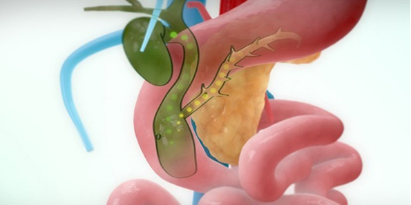
Endoscopic duodenal biopsies play a major role in diseases of the ampulla of Vater and the lower part of the common bile duct. However, there are many cases of severe narrowing or lesions located high in the liver hilum, making the endoscope inaccessible.
Percutaneous intrahepatic biliary biopsy was first introduced in 1980, so far has been widely used worldwide. This technique is performed by inserting a biopsy instrument into the biliary tract through the liver parenchyma. The rate of obtaining successful specimens is quite high, helping to supplement those cases that cannot be biopsied through endoscopic or transhepatic.
Meanwhile, the system of digital subtraction angiography (DSA - abbreviated as "Digital Subtraction Angiography") is an imaging method. Background erasing digital angiography technique combines X-ray and digital image processing, which helps to capture the vascular system in the body and intervene in treatment. The outstanding advantage of this method is that it is minimally invasive and does not require open surgery.

2. When is an intraluminal biopsy necessary?
2.1. Point
Intra-/extrahepatic cholangiocarcinoma, primary/recurrent; Cancer of the stomach, pancreas, or gallbladder with invasion of the biliary tract; Cancer metastatic biliary tract, including: Cancer of the cervix, cancer of the lymphatic system, bronchi; Chronic sclerosing cholangitis; Primary biliary fibrosis.
2.2. Contraindications
Coagulation time INR > 1.5; Platelet count < 50 G/l (need platelet transfusion before intervention); Coagulation factor Prothrombin < 50% (fresh plasma infusion before intervention). Other contraindications include:
Acute liver and biliary tract infections (liver or biliary tract abscess); Bile duct bleeding.
3. Prepare
3.1. Executor
3.2. Vehicle
3.3. Medicine
3.4. General medical supplies
3.5. Special medical supplies
3.6. Patient

3.7. Test form
4. Procedure
4.1. Percutaneous cholangiography
4.2. Biopsy
4.3. Reset percutaneous biliary drainage
5. Complications and treatment
Biliary tract bleeding Requires medical treatment, combined with clinical monitoring, stool, mucosa, and red blood cell volume of the patient. . If biliary tract bleeding persists, consultation with an interventional radiologist is required. Management may be replacement of the drainage catheter or consideration of embolization.
Biliary perforation This condition usually resolves on its own. Very rare complications of biliary peritonitis, if signs are present, consult a surgical specialist.
Biopsy in the lumen of the biliary tract under the guidance of digital scanning of the background brings many advantages, providing information for diagnosis and treatment support. Because of the view into the biliary lumen, the technique allows the diagnosis of biliary tract lesions. Biliary tract diseases, if not treated in time, will cause many dangerous complications, so people need to go to a reputable hospital for timely treatment when there are signs, avoiding complications that affect their health. .
DSA background digitization and percutaneous intraluminal biopsies have been widely used. At Vinmec International General Hospital, there is a team of experienced specialists, with modern equipment, using digital angiography to erase the DSA background in biliary tract biopsies, helping doctors in Diagnosis and treatment are quick and accurate.
Before taking a job at Vinmec Central Park International Hospital from December 2017, Doctor Dang Manh Cuong has over 18 years of experience in the field of ultrasound - diagnostic imaging in Transport Hospitals. Hai Phong, MRI Department of Nguyen Tri Phuong Hospital and Diagnostic Imaging Department of Becamex International Hospital.
For examination and treatment at Vinmec International General Hospital, please come directly to Vinmec Health System or register online HERE.
Dịch vụ từ Vinmec
-
 Nội soi đặt stent điều trị tắc đường mật do sỏi
Phương pháp nội soi mật tụy ngược dòng ra đời đã tạo ra một bước đột phá lớn trong quản lý các bệnh lý đường mật, cho phép can thiệp trực tiếp vào đường mật để chẩn đoán cũng như ...
Đọc thêm -
 Ung thư đường mật giai đoạn cuối phải dùng máy thở có hy vọng cứu chữa không?
Bác sĩ cho tôi hỏi: Cha tôi bị ung thư đường mật di căn gan và hạch, giai đoạn cuối. Hiện nay cha tôi còn bị nhiễm trùng đường ruột, đang được hỗ trợ bằng máy thở, thuốc tăng huyết ...
Đọc thêm -
 Sinh thiết đường mật qua da dưới X quang tăng sáng
Sinh thiết đường mật qua da dưới X quang tăng sáng đóng vai trò quan trọng trong việc xác định chính xác bệnh lý đường mật mà không cần xâm lấn rộng.
Đọc thêm -
 Sốt liên tục sau mổ điều trị ung thư đường mật có nguy hiểm không?
Em bị ung thư đường mật phải cắt bỏ gan trái cùng túi mật. Cắt được 20 ngày thì đau và sốt liên tục. Vậy bác sĩ cho em hỏi sốt liên tục sau mổ điều trị ung thư đường ...
Đọc thêm -
 Dự phòng viêm xơ đường mật
Viêm xơ đường mật là một bệnh lý mãn tính, diễn tiến chậm, đôi khi không có triệu chứng. Người bị viêm xơ đường mật kéo dài có thể dẫn đến tổn thương gan, suy chức năng gan do xơ ...
Đọc thêm