Capture and place a digital biliary stent to erase the DSA . background
This article is professionally consulted by Master, Doctor Vu Huy Hoang - Radiologist - Department of Diagnostic Imaging and Nuclear Medicine - Vinmec Times City International Hospital. Doctor Vu Huy Hoang has 10 years of experience working in the field of diagnostic imaging.
Cholestasis is a condition in which the secretion of bile is blocked, causing bile to enter the bloodstream, causing jaundice and mucous membranes. Background DSA digitized biliary stenting and angiography is a highly advanced technique used in the diagnosis and treatment of obstructions occurring in the bile ducts.
1. What is the technique of taking and placing a biliary stent?
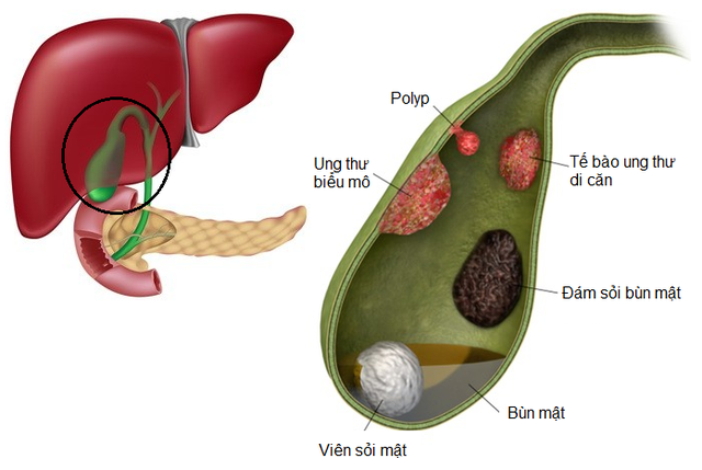
Common causes of malignant biliary obstruction include: pancreatic head cancer, cholangiocarcinoma, gallbladder cancer, and metastatic lesions to the liver hilum. At that time, the patient had 2 lesions, cancer and biliary obstruction. In which, biliary obstruction is often the main cause of death. To solve this problem, since the 1970s, people have carried out percutaneous biliary interventions to release the biliary tree. A biliary stent is a small, thin tube that is used to support the constricted portion of the bile duct. Biliary stenting is a minimal but highly effective intervention, with few complications, and a fast recovery time.
2. Indications and contraindications of the method
2.1 Percutaneous transhepatic cholangiography (PTC)
Percutaneous cholangiography is the first step in percutaneous biliary interventions such as drainage, percutaneous biliary stenting.
This technique is indicated in the following cases:
Differential diagnosis of biliary obstruction that can be treated by surgery or cannot be surgically intervened with medically treated lesions. After failure of endoscopic cholangiography or assessment of complications following endoscopic cholangiography. Evaluation of preoperative biliary stricture lesions (preoperative billan). Contraindicated in patients with:
Coagulation disorders because of the risk of bleeding. Treatment to improve coagulation is required prior to interventions. There is a history of allergy to contrast agents. It is possible to do prophylactic treatment before use with antihistamines and corticosteroids. Abdominal effusion is large, in this case, endoscopic biliary intervention is recommended. In the event of failure of laparoscopic intervention, percutaneous therapy can be performed after drainage of the abdominal fluid. There is no way to enter through the liver because the pleura is entangled or the loops of intestine are too high.

Bệnh nhân rối loạn đông máu chống chỉ định thực hiện
2.2 Percutaneous Transhepatic biliary drainage (PTBD)
Indications of the procedure:
Treatment of obstructive jaundice in case of failure of endoscopic therapy or no indication for endoscopic treatment. Treatment of infectious complications of biliary obstruction such as cholangitis or septic shock. Treatment of biliary obstruction before surgery. Bile drainage (prophylactic treatment) in cases of biliary interventions such as biopsy of intraluminal tumors, biliary stenting. Contraindicated in the following cases:
All contraindications stated in percutaneous cholangiography. Patients with biliary obstruction of multiple sites or only localized obstruction of one segment (usually due to liver metastases).
2.3 Percutaneous transhepatic biliary stenting (PTBS)
Percutaneous biliary stenting is indicated in the treatment of cases of malignant biliary obstruction that cannot be operated on due to late-stage tumor lesions, old age or other comorbidities (diabetes mellitus, chronic obstructive pulmonary disease). Cardiovascular...)
Contraindicated in severe infections (in these cases, biliary drainage should be done first to stabilize treatment), when the diagnosis of malignant biliary strictures is not clear, the lesions remain It is possible to surgically remove the lesion completely.
Contraindicated in severe infections (in these cases, biliary drainage should be done first to stabilize treatment), when the diagnosis of malignant biliary strictures is not clear, the lesions remain It is possible to surgically remove the lesion completely.

Đặt stent đường mật qua da (PTBS) thường được sử dụng trong điều trị tắc đường mật ác tính.
3. How is the radiograph and stent placement of the lower bile ducts performed?
3.1 Percutaneous cholangiography
The technique is performed under aseptic conditions. Biliary puncture under ultrasound guidance or only using a DSA machine Perform cholangiography in appropriate positions to diagnose and reveal the location of the lesion.
3.2 Biliary drainage
Biliary needle puncture (usually Angiocath16-18F) into the biliary tree under ultrasound guidance. Then cholangiography to evaluate the intrahepatic and extrahepatic biliary tree system, the location of biliary obstruction. Insert the catheter into the biliary tract then replace with Amplatzer. Insert a catheter to drain the biliary tract or bring the tip of the catheter down into the duodenum (internal-external drainage).
3.3 Biliary stenting
The technique is the same as for biliary drainage.
After the amplatzer is inserted into the biliary system. Perform angioplasty and insert a stent holder 8-10 mm in diameter through the narrowing site. Prophylactic biliary catheters can be placed into the biliary system depending on whether there is bleeding during the intervention or not. This drainage catheter will be clamped in 24 hours. If there are no complications (the patient has no pain, no fever), the prophylactic catheter can be withdrawn after 48 hours. After the surgical intervention, with percutaneous cholangiography, patients will be monitored at the bedside for about 2-3 hours. If an infection is suspected, antibiotic treatment will be prescribed for 3-5 days. As for the patient undergoing biliary drainage and percutaneous biliary stenting, after the intervention, the patient must be closely monitored next to the intervention room. Depending on the patient's health condition, the patient will be taken to the intensive care room or returned to the hospital bed and continue to take antibiotics after 3-5 days, and at the same time give fluids to replenish the lost bile.
After the amplatzer is inserted into the biliary system. Perform angioplasty and insert a stent holder 8-10 mm in diameter through the narrowing site. Prophylactic biliary catheters can be placed into the biliary system depending on whether there is bleeding during the intervention or not. This drainage catheter will be clamped in 24 hours. If there are no complications (the patient has no pain, no fever), the prophylactic catheter can be withdrawn after 48 hours. After the surgical intervention, with percutaneous cholangiography, patients will be monitored at the bedside for about 2-3 hours. If an infection is suspected, antibiotic treatment will be prescribed for 3-5 days. As for the patient undergoing biliary drainage and percutaneous biliary stenting, after the intervention, the patient must be closely monitored next to the intervention room. Depending on the patient's health condition, the patient will be taken to the intensive care room or returned to the hospital bed and continue to take antibiotics after 3-5 days, and at the same time give fluids to replenish the lost bile.

Bệnh nhân được chỉ định dùng kháng sinh nếu nghi ngờ nhiễm khuẩn
4. Complications may be encountered after the intervention
All procedures, although minimally invasive, still have the risk of complications, with digital biliary stenting and imaging techniques to erase the background of possible complications after intervention such as:
Septic shock. Peritonitis . Bleeding in the abdomen. Pneumothorax.
Septic shock. Peritonitis . Bleeding in the abdomen. Pneumothorax.

Tràn khí màng phổi có thể xảy ra sau khi can thiệp
Depending on the type of complication will be treated according to specific regimens.
Scanning and placing digitalized biliary stents to erase the background is a difficult imaging technique, requires a professional performer, and at the same time, this technique must be performed at adequate medical facilities. facilities, modern machinery system, standard photography room. Therefore, to achieve accurate imaging results, patients need to choose reputable addresses that have DSA angiography machines and have modern and standard medical equipment to perform, from which to have treatment directions. timely illness.
Vinmec International General Hospital is a hospital with a full convergence of general and specialized doctors to perform, examine, operate, diagnose and treat diseases. In particular, Vinmec has been and continues to invest in modern machinery systems such as ultrasound machines, computerized tomography, magnetic resonance imaging to serve the examination and treatment of diseases.
Besides, Vinmec has also performed DSA interventional angiography techniques in many cases to promptly provide results and treatment directions for many customers.
Để đặt lịch khám tại viện, Quý khách vui lòng bấm số HOTLINE hoặc đặt lịch trực tiếp TẠI ĐÂY. Tải và đặt lịch khám tự động trên ứng dụng MyVinmec để quản lý, theo dõi lịch và đặt hẹn mọi lúc mọi nơi ngay trên ứng dụng.


