Impetigo: What you need to know
Article written by Specialist Doctor I Tran Van Sang - Department of Medical Examination & Internal Medicine - Vinmec Danang International Hospital
Impetigo is a common superficial bacterial infection of the skin, characterized by vesicles, vesicles that rapidly turn pus, then rupture leaving shallow erosions and honey-colored scabs. The term impetinisation is used to refer to superficial infections secondary to a wound or certain skin condition. When the lesion is deeply ulcerated, it is called an impetigo.
1. Classification of impetigo
Impetigo is common in children, more boys than girls. In adults, rarely, the disease occurs in people with weakened immunity. The disease is common in the summer, common in developing countries, with unsanitary living conditions and a large population. Impetigo is common after a number of skin diseases such as atopic dermatitis, scabies, chickenpox, insect stings, thermal burns, dermatitis.
Classification of impetigo, including 3 types:
Bullous Impetigo Non-bullous Impetigo Impetigo

Chốc thường xuất hiện ở các bé nam
2. Causes of disease
The cause of impetigo is staphylococcus aureus or streptococcus bacteria. And sometimes a combination of these two types of bacteria. Nonbullous impetigo can be caused by group A beta-hemolytic streptococcus, staphylococci, and/or streptococcus entering small wounds in the skin where proteins help the bacteria attach organization. Bullous impetigo is usually caused by the exfoliative staphylococcal toxin (exfoliatin A-D) acting on the desmoglein 1 bridge of the epidermal squamous cells, dissecting the superficial layer of the epidermis, creating a pemphigus-like appearance. leaf scales. Impetigo is usually caused by streptococcus but can be combined with Staphylococcus aureus, occurs in immunocompromised, the elderly, people with chronic diseases.
3. Clinical symptoms
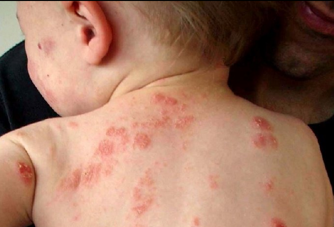
Impetigo is common in open skin such as the face, around the natural cavities of the mouth and nose, on the scalp, hands and feet, but also on the trunk and other parts of the body. The disease presents with a single lesion or multiple lesions. Patients may have fever, fatigue, and swollen glands. The non-bullous moment usually begins as a pink macula, progresses to vesicles, rapidly turns pus, quickly ruptures, leaving scaly, honey-yellow scabs. When the scabs fall off, the skin is red and moist, and when it heals, it leaves a dark macule. If left untreated, the disease can heal on its own in 2-4 weeks without scarring. Lesions can spread to other areas by self-infection, by scratching.
Any area of the body can be affected, but the face and extremities are most commonly affected. Lesions may be mildly pruritic or asymptomatic, with or without a surrounding red halo. Peripheral lymph nodes are usually enlarged. Patients may have minor trauma, insect stings, scabies, chickenpox, atopic dermatitis at the impetigo site.
Impetigo starts as impetigo without blisters but progresses to mid-concave necrotic ulcers, slow to heal, leaving scars. Bullous impetigo begins with small blisters that gradually grow into blisters. The blisters are shallow, fragile, small or large, containing a clear, yellow fluid, then turn dark yellow, rupture in 1 to 3 days, leaving a thin skin border around a moist red patch that heals without scarring. There may or may not be a red halo around the blister. Lesions are common on the face, trunk, extremities, buttocks, then spread to the distal ends by self-infection. Unlike non-bullous impetigo, bullous impetigo can have lesions on the cheek mucosa, is less contagious, and regional lymph nodes are not enlarged.

Chốc bọng nước khởi phát với những mụn nước nhỏ
4. Complications of impetigo
Glomerulonephritis Cellulitis Scarlet fever
5. Diagnosis of impetigo
Diagnosis of impetigo is mainly based on clinical symptoms such as vesicles, blisters that turn pus rapidly, break to form a shallow slide on the skin, crusted secretions, honey yellow color, common locations are: around the nose, mouth, scalp, hands and feet..
In addition, some tests can be done such as staining for bacteria, bacterial culture, white blood cell formula (with neutrophilic), diseased tissue learn.
Histopathological features of impetigo:
Impetigo without blisters: with gram-positive staphylococci, pustules containing neutrophils in the epidermis, dense inflammatory infiltrates in the superficial dermis. Bullous impetigo: the epidermis is split in the granulation layer without inflammation, no bacteria, cleavage, mild inflammatory infiltrate in the superficial dermis.– Impetigo: deep ulcers, with catching cocci gram color in the dermis.

Chẩn đoán bệnh chốc dựa vào biểu hiện lâm sàng hoặc thông qua các xét nghiệm
6. Treatment of impetigo
Follow these steps:
Wash the lesion, gently remove the exudate. Use antiseptics: Apply Milian, methyl methyl blue.. Or topical antibiotic ointment (fusidic acid 2%, mupirocin 2%) ... If impetigo spreads, systemic antibiotics can be used such as: Augmentin, Cefaclor, Azithromycin...
When there are severe symptoms such as high fever and complications, hospitalization should be given.
=>> SEE ALSO: What drugs are used for impetigo?
7. Prevention of impetigo
Clean body Use antibacterial shower gel daily like Cetaphil, Lactacyd Use separate towels Change clothes and wash daily Treat sources of infection Prevent spreading disease to others:
Avoid close contact with Others Keep the child out of school until the scabs have dried
Để đặt lịch khám tại viện, Quý khách vui lòng bấm số HOTLINE hoặc đặt lịch trực tiếp TẠI ĐÂY. Tải và đặt lịch khám tự động trên ứng dụng MyVinmec để quản lý, theo dõi lịch và đặt hẹn mọi lúc mọi nơi ngay trên ứng dụng.


