
Lung/pleural biopsy under computed tomography
1. What is lung biopsy technique, pleural biopsy under computed tomography?
Conventional imaging techniques such as chest X-ray and computed tomography (CT) scan of the chest can help detect lung lesions, but to confirm the nature of the lesion, images are often not enough. Using a needle for a CT-guided transthoracic biopsy provides more information for diagnosis. This is a safe, minimally invasive technique that gives accurate diagnostic results.
Lung / pleural biopsy under the guidance of computed tomography is often indicated in cases of lung, pleural and mediastinal lesions that need to confirm the diagnosis, suspect tumor lesions, inflammation, or tuberculosis. ..
2. What to prepare before performing lung biopsy, pleural biopsy under computed tomography?
2.1. The person performing the trick
2.2. Means of performing CT-guided transthoracic biopsy
Computerized tomography machine; film, film printer, image storage system Local anesthetic, general anesthetic (if there is an indication for anesthesia), water-soluble iodinated contrast agent, skin antiseptic solution Biopsy needle specialized, needle pump, cotton, gauze,...
2.3. Prepare patient for CT-guided transthoracic biopsy
Patients will be carried out a number of important tests such as blood count test, complete coagulation test, echocardiogram, ECG, respiratory function measurement, ... to ensure health conditions. suitable for performing biopsies.
The medical staff will instruct the patient to fast for 4 hours before the lung biopsy / pleural biopsy. The patient must inform the doctor of all the medicines they are taking, the drugs that are allergic,... The doctor will ask to stop the anticoagulants, antiplatelet drugs (such as Heparin, Aspirin, etc.) Wafarin,...) a few days before the procedure.
Female patient, if pregnant or suspected to be pregnant, must notify the doctor or technician in the imaging room. To avoid radiation exposure to the fetus, imaging methods are not usually performed. However, where it is necessary to proceed, precautions will be taken to minimize the potential for fetal exposure. Patients should also inform their doctor of other illnesses or health conditions, if any.

3. How is a lung/pleural biopsy under computed tomography done?
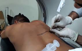
Localized CT scan for the patient to locate the lesion. After determining the location, the skin belonging to this location and the surrounding skin will be disinfected and cleaned. A towel that has been sterile and has a circular opening is placed over the disinfected skin area.
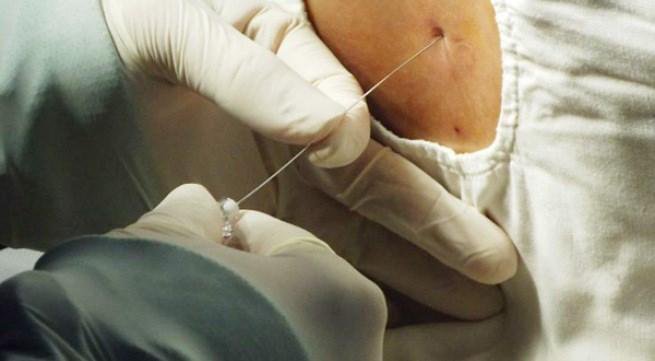
Apply local anesthetic around the area to be examined. The doctor will make a very small incision in the skin with a surgical blade, then insert a guide needle through the skin incision site under the guidance of computed tomography to bring the needle close to the site of the lesion (tumor). ).
A specialized biopsy needle will be passed through the guide needle to reach the tumor. The doctor will conduct a biopsy cutting into the tumor, taking 2-3 samples from different directions. Because when the patient breathes, the tumor will move with the breathing, so the patient should try to hold their breath while the needle is inserted through the pleura, lung parenchyma and during the biopsy.
The team takes several cuts through the biopsy area to check if the patient has conditions such as bleeding in the lungs, pleura, pneumothorax,... or not.
The procedure is usually completed within an hour. The patient is instructed to stay in bed for at least 6 hours, the pulse, blood pressure, and general condition of the patient will be closely monitored for 24 hours after the procedure.
4. Possible complications in lung/pleural biopsy under computed tomography
4.1. Complications of pneumothorax

4.2. Accident bleeding into the mouth, nose; coughing up blood during lung biopsy, pleural biopsy
Some other possible complications such as bleeding lung parenchyma, severe pleura, chest pain, damage to the diaphragm and organs under the diaphragm, chest wall, ... The doctor will treat depending on symptoms and specific causes. Therefore, you should choose a lung biopsy at a large, reputable medical facility not only in the hospital system in general but also stand out and be a leader in the field of lung cancer diagnosis and treatment.
Để đặt lịch khám tại viện, Quý khách vui lòng bấm số HOTLINE hoặc đặt lịch trực tiếp TẠI ĐÂY. Tải và đặt lịch khám tự động trên ứng dụng MyVinmec để quản lý, theo dõi lịch và đặt hẹn mọi lúc mọi nơi ngay trên ứng dụng.
Dịch vụ từ Vinmec
-
 Bị u phổi di căn sang xương nên điều trị thế nào cho hợp lý?
Tôi mổ thay khớp háng tháng 11/2019, sau đó về điều trị ở nhà. Tuy nhiên tôi thấy đau nhiều ở cả hai chân, tôi đi khám tổng thể thì bác sĩ thấy u ở phổi và kết luận bị ...
Đọc thêm -
 Bác sĩ chẩn đoán ung thư phổi như thế nào?
Dựa trên cách nhìn của tế bào ung thư dưới kính hiển vi, bác sĩ phân thành 2 loại ung thư phổi, đó là ung thư phổi tế bào nhỏ và ung thư phổi không phải tế bào nhỏ. Nếu ...
Đọc thêm -
 Những điều cần biết khi bạn mắc ung thư phổi không tế bào nhỏ (non-small cell lung cancer) - Phần 2
Ung thư có thể bắt đầu từ bất cứ nơi nào trong cơ thể. Ung thư bắt đầu ở phổi gọi là ung thư phổi. Nó bắt đầu khi các tế bào trong phổi phát triển ngoài tầm kiểm soát ...
Đọc thêm -
 Đau sau vai kèm hơi thở ngắn sau sinh thiết phổi có sao không?
Em đã sinh thiết phổi được 1 tuần. Sau ngày sinh thiết em đã chụp X quang để kiểm tra thì không thấy tràn dịch hay tràn khí màng phổi, nhưng đến bây giờ là 1 tuần em vẫn còn ...
Đọc thêm -
 Viêm màng phổi có dịch có nguy hiểm không?
Ba em hay ho và đau tức ngực. Sau khi xét nghiệm và chọc hút dịch thì bác sĩ không kết luận được gì. Bác sĩ có thể cho em biết ba em bị gì được không? Trên phiếu xét ...
Đọc thêm