
Percutaneous biliary biopsies under enhanced X-ray
1. An overview of biliary tract cancer
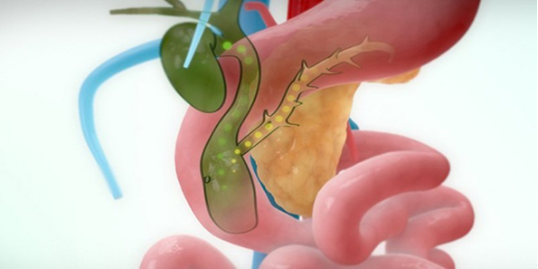
intrahepatic cholangiocarcinoma; extrahepatic cholangiocarcinoma. When cells in the bile ducts grow abnormally or develop into malignancies, they increase in size and invade surrounding structures and other parts of the body.
In the early stages, cholangiocarcinoma often has no specific warning signs. Only when the cancer cells grow will the patient have the following symptoms:
Yellow eyes and skin Tea-colored urine Upper stomach or back pain Pale stools Nausea or vomiting Poor appetite, weight loss The above symptoms also occur in many other common diseases, so the patient needs to go to the hospital for examination and examination for accurate results.

2. Diagnostic measures for biliary tract cancer
Percutaneous bile duct biopsies under enhanced radiography play an important role in accurately identifying biliary tract disease without extensive invasiveness.
3. Indications and contraindications
Cases of benign or malignant biliary obstruction but the cause cannot be determined by endoscopic or transhepatic biopsy. Chronic sclerosing cholangitis. Primary sclerosing cholangitis. Pancreatic cancer, gastric cancer, gallbladder cancer invade biliary tract. Metastatic biliary tract cancer (bronchial cancer, lymphoma, cervical cancer). Intra and extrahepatic cholangiocarcinoma: Recurrent or primary. Contraindications:
Patients with severe coagulopathy (consider): INR >1.5; platelet count <50 G/l: platelet transfusion is required before intervention; Prothrombin ratio <50%: need fresh plasma before intervention. Other contraindications: Acute liver and biliary tract infections (bile duct abscess, liver abscess). Bleeding of the biliary tract

4. Percutaneous biliary biopsy under enhanced X-ray
4.1 Preparation for the procedure
Implementation team:
Specialist, ancillary doctor; Electro-optical technician; Nursing; Anesthesiologist/technologist (if patient is difficult to cooperate). Means of use:
Fluoroscopy; Film, film printers and image acquisition systems; Lead vest, apron to help shield from X-rays; Flat and curved transducer ultrasound machine; Sterile plastic bag wrapped ultrasound transducer. Medicines:
Includes local anesthetics; General anesthesia (applied as needed); Anticoagulants and anticoagulants neutralizers; Water-soluble iodinated contrast agent; The solution helps to disinfect the skin and mucous membranes. Common medical supplies:
5.10ml syringe; Distilled water (physiological saline); Surgical clothing; Aseptic interventional kits (knife, scissors, tongs, tool tray, etc.); Medicine box and accident first aid box; Cotton gauze, medical bandages for surgery. Special medical supplies:
Chiba cholangiography needle and corresponding guide wire; Set of tubes into the vessel lumen; 4-5F angiography catheter; Standard conductor 0.035''; Endoscopic biopsy forceps; Drainage catheter; Suture to fix the catheter. Patients need to prepare:
Be explained in detail about the procedure and administrative procedures before proceeding. Examination and clinical examination before the procedure. Fasting, drinking before 6 hours. Do not drink more than 50ml of water. In the intervention room: the patient lies on his back. The doctor installs a machine to monitor breathing, pulse, electrocardiogram, blood pressure, and oxygen saturation in the peripheral blood. Disinfect and cover with a sterile towel with holes. In case the patient is easily excited, unable to lie still, a sedative will be prescribed.
4.2 Percutaneous biliary tract biopsy procedure
Step 1: Percutaneous cholangiography
Check percutaneous biliary ducts with contrast. Push the wire in and withdraw the bile duct. Through the guide of the guide wire, push the catheter into the biliary tree and capture the biliary tree through the catheter. Determine the expected biopsy site. Step 2: Biopsy of the biliary tract
Remove the catheter, place the set of tubes in the lumen and enter the biliary tree through the wire. Withdraw the lead, then use biopsy pliers to cut into the lumen of the bile duct through the tube into the lumen. Cut about 3-5 pieces of tissue in the bile duct for testing. Step 3: Reinsert percutaneous biliary drainage
Withdraw the biopsy forceps from the biliary tract and the duct. Insert the guide wire into the biliary lumen and withdraw the tube into the lumen Reestablish the percutaneous biliary drainage catheter.

5. Complications after the procedure and treatment direction
Bleeding: Monitor for clinical signs. stool, mucosa, erythrocyte volume index) combined with medical treatment. If biliary bleeding persists, consult a physician. The drains can be replaced or the plugs re-examined. Biliary perforation: Usually resolves spontaneously. Rarely, peritonitis complications, if any, require surgical consultation. Vinmec International General Hospital with a system of modern facilities, medical equipment and a team of experts and doctors with many years of experience in medical examination and treatment, patients can rest assured to visit. and hospital treatment.
Before taking a job at Vinmec Central Park International Hospital from December 2017, Doctor Dang Manh Cuong has over 18 years of experience in the field of ultrasound - diagnostic imaging in Transport Hospitals. Hai Phong, MRI Department of Nguyen Tri Phuong Hospital and Diagnostic Imaging Department of Becamex International Hospital.
To register for examination and treatment at Vinmec International General Hospital, you can contact Vinmec Health System nationwide, or register online HERE.
Dịch vụ từ Vinmec
-
 Ung thư đường mật giai đoạn cuối phải dùng máy thở có hy vọng cứu chữa không?
Bác sĩ cho tôi hỏi: Cha tôi bị ung thư đường mật di căn gan và hạch, giai đoạn cuối. Hiện nay cha tôi còn bị nhiễm trùng đường ruột, đang được hỗ trợ bằng máy thở, thuốc tăng huyết ...
Đọc thêm -
 Có thể chẩn đoán ung thư đường mật qua chỉ số creatinin máu không?
Chào bác sĩ. Ba em được chẩn đoán là mắc K đường mật, nhưng khi xét nghiệm chỉ số creatinin máu có kết quả là 40-120 umol/l. Vậy ba em có bị vấn đề gì không ạ? Em xin cám ...
Đọc thêm -
 Sốt liên tục sau mổ điều trị ung thư đường mật có nguy hiểm không?
Em bị ung thư đường mật phải cắt bỏ gan trái cùng túi mật. Cắt được 20 ngày thì đau và sốt liên tục. Vậy bác sĩ cho em hỏi sốt liên tục sau mổ điều trị ung thư đường ...
Đọc thêm -
 Biến chứng và điều trị sỏi đường mật trong gan
Sỏi đường mật trong gan là bệnh lý khá phổ biến, thường để lại các biến chứng nguy hiểm như nhiễm trùng đường mật, viêm gan, xơ gan, ung thư đường mật trong gan, nhiễm trùng huyết,... Việc phát hiện ...
Đọc thêm -
 Chụp số hóa xóa nền và sinh thiết trong lòng đường mật qua da
Sinh thiết trong lòng đường mật dưới hướng dẫn của chụp số hóa xóa nền là một trong những kỹ thuật hiện đại. Phương pháp này giúp các bác sĩ can thiệp trực tiếp tổn thương trong lòng đường mật ...
Đọc thêm