
When to perform routine joint radiographs?
Article written by Dr. Pham Quoc Thanh - Diagnostic Imaging Department - Vinmec Hai Phong International General Hospital
Routine joint radiography is an in-depth investigation, using contrast material (iodine or gas-containing contrast) directly into the joints under the monitoring and imaging of an X-ray machine with an enhanced fluorescence screen. .
1. What is routine joint X-ray?
X-ray is an imaging technique that helps doctors diagnose certain medical conditions. The X-ray machine emits X-rays that penetrate the organs (dense tissues in the body such as bones,... or less dense tissues such as muscles,...) at a sufficient dose and create clear images. outline the organs inside the body. This is an advanced method of medicine, often indicated for implementation, especially for problems related to bones and joints.
Routine joint radiography is an in-depth investigation, using contrast material (iodine or gas-containing contrast) directly into the joints under the monitoring and imaging of an X-ray machine with an enhanced fluorescence screen. .
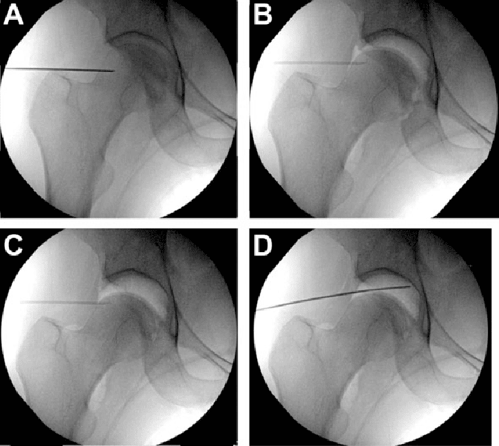
X-ray with bright fluorescent screen helps the radiologist to see the movement of the internal organs. When iodinated contrast agent is injected into the joint, it will evenly coat the inner surface of the joint structures, showing white light to help the radiologist understand the anatomical structures and activities of the joints.
Currently, images are stored in digital form, which can be easily processed, reviewed and especially compared with each other for diagnosis and treatment.

2. When to perform routine joint X-ray?
X-ray images of joints help doctors evaluate changes in the structure and function of joints and help determine treatment options: medical, surgical or joint replacement.
Arthroscopy is indicated for unexplained persistent pain and discomfort in joints.
This survey is often used to detect abnormalities in the joints:
Shoulders Wrists Knees Ankles
3. How does routine joint X-ray work?
3.1 Preparation The patient does not need to be hospitalized, fasting or fasting is not required.
The patient will fill out a questionnaire and consent form before the procedure:
Current medications (list of drugs), allergies if any, especially to iodinated contrast if previously here used.

The patient should also report the most recent medical condition. Change into a robe and remove any jewelry and metal items you carry. Female patients should notify their physician or technician if pregnancy is suspected. Some imaging tests are not done during pregnancy because radiation can affect the fetus. In cases where it is imperative to proceed, a number of safety protection measures must be taken, minimizing radiation contamination to the fetus. 3.2 Implementation Step 1: Technician prepares supplies, injection tools, and scanner.
Step 2: The patient is placed on the X-ray table. Take some dimensions, the position of the joint before injection (to compare with the image after the contrast material is injected).
Step 3: Carry out the technique:
Disinfect the skin around the joint several times. Local anesthetic may be injected (if needed) The radiologist will insert a thin needle, of the required length, through the skin and straight into the joint space. Inject contrast material (or gas) into the joint. The patient may feel a stretch in the joint as the contrast agent is injected. After the needle is removed, ask the patient to move the joint gently so that the contrast agent is evenly placed in the joint. Take the same scan as step 2. The survey lasts about 20 - 30 minutes. Note: Joint CT or Magnetic Resonance Imaging may be performed immediately after radiographic positions for a more accurate assessment of the structures within the joint.
Step 4: Finish the procedure, apply pressure to the needle puncture site, monitor the patient and wait for the results.
4. Complications that may occur with routine joint X-ray
Any intervention performed on the human body, even under the most safe conditions, carries the risk of complications:
Because the contrast agent is injected directly into the joint, the reaction Allergic reactions are very rare (mild can cause nausea, severe can still have cardiovascular complications). Risk of infection when the needle is inserted into the joint. Those performing the procedure always take the utmost care to control infection to avoid this risk. After the procedure, the joint may be sore and slightly swollen. Patients can apply ice to the joint to reduce swelling and pain. Common pain relievers can help improve pain. The above symptoms should disappear after 48 hours. If it lasts longer, the patient should immediately notify the doctor to choose another treatment. Joint movement should be limited for 24 to 48 hours after the scan. For detailed advice, please come directly to Vinmec health system or register online HERE.
Role of radiographs in the diagnosis and treatment of shoulder periarthritis Purpose of TMJ radiographs Role of radiographs in diagnosing knee arthropathy
Dịch vụ từ Vinmec
-
 Quy trình chụp cắt lớp vi tính cột sống cổ có tiêm thuốc đối quang
Ngoài phương pháp chụp X-quang, chụp CLVT cột sống cổ có tiêm thuốc đối quang cũng là kỹ thuật chẩn đoán hình ảnh có độ chính xác cao và rất an toàn, giúp bác sĩ có thể phát hiện được ...
Đọc thêm -
 Quy trình chụp cộng hưởng từ tĩnh mạch có tiêm thuốc đối quang từ
Cộng hưởng từ tĩnh mạch có tiêm thuốc đối quang từ thường được sử dụng trong chẩn đoán và theo dõi những bệnh lý về đường tĩnh mạch như thông động tĩnh mạch, dị dạng hay huyết khối. Là một ...
Đọc thêm -
 Tiếng kêu trong khớp sau khi đóng đinh nội tủy xương chày có sao không?
Em năm nay 29 tuổi, em bị tai nạn giao thông gãy cả xương chày và xương mác. Gãy 1/3 xương hở độ 1. Xương gãy vẫn nằm kín dưới da. Em đã được phẫu thuật đóng đinh nội tủy ...
Đọc thêm -
 Gãy xương chày 4 năm bị lệch có hồi phục được không?
Em bị tai nạn gãy 2 xương chân phải và được bó bột, cứ hơn 1 tháng là thay bột, đến đây đã hơn 4 năm mà mà chân em chạy vẫn cà nhót và em đi khám và chụp ...
Đọc thêm -
 Chỗ bị va đập ở đầu mềm liệu có phải chụp X quang không?
Bác sĩ cho em hỏi, bé nhỏ em được 26 ngày, bị bé lớn 2 tuổi lấy đồ chơi bằng nhựa (cứng) đập vào đầu bé nhỏ, gần ngay thóp bé nhỏ, không chảy máu, nhưng có bị mềm khu ...
Đọc thêm