
How are moles formed?
1. Moles and their location on skin structure
1.1 The epidermis The epidermis is the top thin layer of skin that is visible to the naked eye. It acts as a waterproof barrier protecting the body from bacteria and other microscopic organisms.
Cells in the epidermis:
Keratinocytes are specialized skin cells that make up the different layers of the epidermis. The outermost part of the epidermis (the stratum corneum) is made up of keratinocytes that have lost their nucleus, which contain a protein called keratin that makes them stiff and bound together to form a water-repellent barrier. Keratinocytes are continuously shed and replaced by squamous cells (which turn into keratinocytes during migration to the top of the skin).
Squamous cells are living keratinocytes that produce keratin, an important protein for the skin.
Keratinocytes are small, round cells that are the only cells in the epidermis that divide to produce new skin cells, replacing those that are dead and shed from the surface of the skin) that make up the bulk of the skin. large basal layer (innermost layer of the epidermis).
Melanocytes are located in the basal layer of the epidermis. They are frequently alternated between keratinocytes. Melanocytes produce melanin, a pigment protein that provides skin color. hair color and protects against the harmful effects of UV radiation.
1.2 Dermis The second layer of skin, thicker and located below the epidermis. Contains blood vessels, lymph, nerve endings, muscle fibers, oil glands, sweat, and hair follicles.
There are 2 small layers in the dermis:
Superficial dermis: Made up of loose connective tissue, blood vessels and nerves. The papillae are like fingers that connect the dermis to the epidermis and provide the epidermis with important nutrients. Deep dermis: A dense network of collagen fibers, the connective tissue that gives the skin its elasticity and strength. It is home to a large number of blood vessels, nerves, as well as lymph vessels, glands and hair follicles. 1.3 Bottom layer (subcutaneous fat layer) Subcutaneous layer or subcutaneous fat layer: A thick layer of fat and connective tissue below the dermis.
Like the dermis, it contains a rich supply of blood and lymph. It insulates and saves body heat, acts as a shock absorber to help protect tissues and organs under the skin from injury, and is a stored energy source.
1.4 Where the mole is on the skin

Most moles appear in childhood and in the first 25 years of life. An adult usually has an average of 10-40 moles.
As the years go by, moles usually change slowly, becoming larger in size and/or changing color. Sometimes, hairs develop in the mole. Some moles do not change, while others may gradually disappear over time.
2. The formation of moles
Melanocytes and Melanin
Melanocytes are cells located in the lower part of the epidermis, just above the dermis. They make a pigment called melanin, which gives color to the skin, hair, and parts of the eyes. Melanin protects the skin from ultraviolet (UV) radiation, the harmful rays from the sun. Mole (cluster of Melanocytes)
A mole is a group of melanocytes that appear as a pigmented spot on the skin. They can be flat or raised, round or oval, and are usually smaller than a pencil eraser.
Although moles are generally benign and unchanged, they can sometimes become cancerous. The first sign of melanoma is usually a change in the size, shape, or color of an existing mole, or the appearance of a new mole in adulthood.
Both light-skinned and dark-skinned people have the same number of melanocytes. The difference in skin color is the result of a difference in the amount of melanin and the size of the melanin packets.
Eumelanin is a reddish-yellow pigment most commonly found in fair-skinned people with red hair. Some redheads don't seem to have any moles. People with a lot of eumelanin are more prone to tanning and are protected from UV radiation. People with less eumelanin are more likely to have freckles or burns. Pheomelanin: The most abundant human melanin, found in brown or black skin and hair. Albinos: Unable to produce a normal amount of melanin and therefore have reduced levels or no pigmentation in the skin, hair and eyes.
3. Types of moles that need attention
Dysplastic nevi are moles that are larger than average (larger than a pencil eraser) and irregularly shaped. They tend to be uneven in color with a dark brown center and lighter, uneven edges. These nevi are more likely to become melanoma. In fact, people with 10 or more dysplastic Nevi-type moles are 12 times more likely to develop melanoma. Any unusual changes to the mole should be checked by a dermatologist to evaluate for skin cancer.

4. How to tell if a mole is cancerous?
It is recommended to check your skin with a mirror or ask someone to help you. Pay special attention to areas of skin that are frequently exposed to the sun, such as the hands, arms, chest, neck, face, ears, legs, and back.
If a mole doesn't change over time, don't worry too much about it. Conversely, if you see any change in an existing mole, appear a new mole, or want to remove it for cosmetic reasons, consult a dermatologist.
The following ABCDEs are important features to consider when examining moles. If a mole has any of the signs listed below, get it checked out by a dermatologist right away. Because it could be cancer.
Asymmetry: One half of the mole does not match the other half. Shape: The border or edge of the mole is torn, blurred, or irregular. Color: The color of the mole is not the same (transparent or with brown, black, blue, white, red pigments). Diameter: The diameter of the mole is larger than a pencil eraser. Growth: The mole is changing in size, shape, or color. Melanoma is a form of skin cancer. The most common locations for melanoma in men are the chest and back and in women the lower legs. Melanoma is the most common cancer in young women.
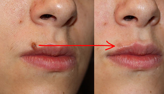
5. How are moles treated?
Depending on the biopsy results, in the absence of malignant cells, the doctor may remove the entire mole or use a laser to remove the mole.
In the case of melanoma, depending on the invasive nature of the tumor, the doctor can surgically remove the tumor or combine with radiation therapy or chemotherapy after surgery.
6. Who is prone to having moles?
7. What factors increase the risk of moles?

Để đặt lịch khám tại viện, Quý khách vui lòng bấm số HOTLINE hoặc đặt lịch trực tiếp TẠI ĐÂY. Tải và đặt lịch khám tự động trên ứng dụng MyVinmec để quản lý, theo dõi lịch và đặt hẹn mọi lúc mọi nơi ngay trên ứng dụng.
Dịch vụ từ Vinmec
-
 Mụn đầu đen để lâu có thành nốt ruồi không?
Mụn đầu đen là tình trạng rất phổ biến, dễ dẫn đến các vấn đề về da khác như mụn bọc, mụn mủ... và có ý kiến cho rằng mụn đầu đen không điều trị sẽ trở thành nốt ruồi ...
Đọc thêm -
 U hắc tố lành tính của da
U hắc tố là một tổn thương xuất hiện ở da. U hắc tố gồm 2 loại là u hắc tố lành tính và u hắc tố ác tính. Bài viết sau đây sẽ trình bày những đặc điểm của ...
Đọc thêm -
 Cách xóa nốt ruồi mà không để lại sẹo?
Chào bác sĩ! Thưa bác sĩ, cháu có một nốt ruồi nổi ở gần mũi. Bác sĩ cho cháu hỏi cách xóa nốt ruồi mà không để lại sẹo ạ? Rất mong bác sĩ tư vấn, cảm ơn bác sĩ!
Đọc thêm -
Hiệu quả của việc làm mờ sẹo sáng da từ chiết xuất táo đỏ
Không chỉ được sử dụng trong các món ăn bổ dưỡng giúp hồi phục, tăng cường sức khỏe, nhiều nghiên cứu mới đây còn chỉ ra rằng những thành phần trong táo đỏ có khả năng làm mờ sẹo, sáng ...
Đọc thêm -
 Thuốc Elastiderm: Công dụng, chỉ định và lưu ý khi dùng
Thuốc Elastiderm dạng kem bôi được xem là sản phẩm làm sáng da có hiệu quả điều trị rất cao, nhờ ức chế các enzyme trong tế bào da. Elastiderm tác dụng chính để tẩy trắng da, làm sáng các ...
Đọc thêm