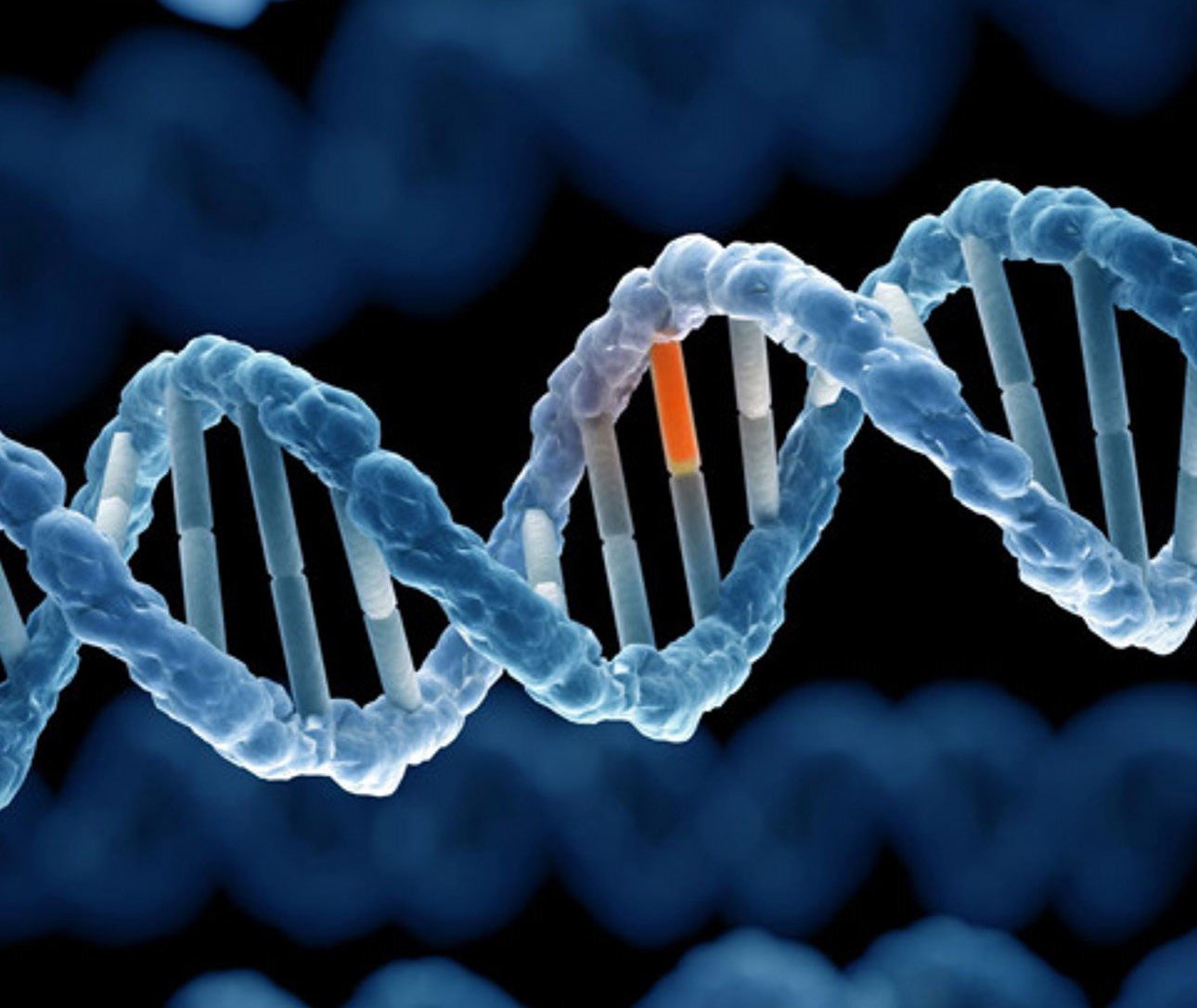
7 genetic tests to do during pregnancy
Prenatal diagnosis is the use of exploratory methods during pregnancy to detect morphological or chromosomal abnormalities of the fetus.
1. AZF . gene test
Analysis of the Y chromosome in men with severe azoospermia or oligozoospermia has resulted in the identification of three regions in the chromosomal portion of the human Y chromosome (Yq11) that are frequently deleted in males unexplained spermatogenesis failure.
PCR analysis of trace elements in the AZFa, AZFb and AZFc (AZF: Azoospermia Factor) regions of the human Y chromosome is an important screening tool in the process of studying infertile men for the selection of infertile men. assisted reproductive technology. Y-chromosomal polymorphisms are the most common genetic cause of male infertility, and screening for these microsomal or heavy-chromosomal men is now standard practice in many infertility centers. .
Devyser AZF v2 kit diagnostic test based on PCR amplification of sequence tagged sites (STS) in the AZFa, AZFb and AZFc regions on the Y chromosome. Successful amplification of the marker STS showed the presence, while absence of PCR amplification was indicative of deletion. Devyser's unique single-tube approach simplifies workflow, reduces practice time, and minimizes the risk of sample mix-ups and contamination during both basic and extensive AZF analysis.
2. Thalassemia gene test
Thalassemia is a group of inherited blood disorders that can be passed from parents to their children and affect the amount and type of hemoglobin the body produces.
Hemoglobin (Hb or Hgb) is a substance found in all red blood cells (RBCs). It is important for the proper function of red blood cells because it carries the oxygen that red blood cells provide throughout the body. A part of hemoglobin called heme is the molecule that has iron in it...
Alpha thalassemia is sometimes confused with iron deficiency anemia because both disorders have red blood cells that are smaller than normal (microcytic). ). If someone has thalassemia, his or her iron levels are not expected to be low. Iron therapy will not help people with alpha thalassemia and can lead to iron overload, which can damage organs over time.
Evaluation of hemoglobin (Hb) disease (hemoglobin electrophoresis). This test evaluates the type and relative amount of hemoglobin present in red blood cells. Hemoglobin A (Hb A), which includes both alpha and beta globin, is the type of hemoglobin that normally makes up 95% to 98% of hemoglobin in adults. Hemoglobin A2 (HbA2) typically makes up 2% to 3% of hemoglobin in adults, while hemoglobin F usually makes up less than 2%.
Beta thalassemia upsets the balance of beta chain formation and alpha hemoglobin and causes an increase in those small hemoglobin components. Therefore, people with beta thalassemia major often have an Hb F ratio. People with beta thalassemia major usually have a higher Hb A2 ratio. Hb H is a less common form of hemoglobin that can be seen in some cases of alpha thalassemia. Hb S is the hemoglobin that is more common in people with sickle cell disease.

Xét nghiệm gen giúp phát hiện bệnh Thalassemia
Evaluation for hemoglobin (Hb) disease is used for state-regulated neonatal hemoglobin screening and prenatal screening when parents are at high risk for hemoglobin abnormalities.
Amniotic fluid genetic testing is used in rare cases where the fetus is at higher risk of thalassemia. This is especially important if both parents are likely to carry the mutation because it increases the risk that their child may inherit a combination of abnormal genes, which causes a more severe form of thalassemia.
3. Genetic testing for hereditary thromboembolic disease
3.1. Factor V Leiden (FV) gene mutation Factor V Leiden thrombophilia is an inherited disorder of blood clotting. Factor V Leiden is the name of a specific gene mutation that leads to thrombophilia, which is an increased tendency to form abnormal blood clots that can block blood vessels.
People with factor V Leiden thrombophilia have a higher than average risk of developing a type of blood clot called deep vein thrombosis (DVT). DVTs occur most often in the legs, although they can also occur in other parts of the body, including the brain, eyes, liver, and kidneys. Thrombotic Factor V Leiden also increases the risk that blood clots will break out of their original place and travel through the bloodstream. These blood clots can take up residence in the lungs, where they are known as pulmonary embolisms. Although factor V Leiden thrombosis increases the risk of blood clots, only about 10% of individuals with a factor V Leiden mutation develop abnormal blood clots.
Factor V Leiden mutation is associated with a slightly increased risk of miscarriage (miscarriage). Women with this mutation are two to three times more likely to have multiple miscarriages (recurrent) or miscarriages in the second or third trimester. Some research suggests that mutated factor V Leiden may also increase the risk of other complications during pregnancy, including pregnancy-induced high blood pressure (pre-eclampsia), fetal growth retardation, and placental abruption. premature separation from the uterine wall (placental abruption). However, an association between factor V Leiden mutations and these complications has yet to be confirmed. Most women with thrombotic factor V Leiden have difficulty having normal pregnancies.

Đột biến gen yếu tố V Leiden là một rối loạn di truyền về đông máu
3.2. Factor II prothrombin G20210 (FII) gene mutation The prothrombin 20210 mutation, also known as a Factor II mutation, is an inherited condition that increases your blood's chance of forming dangerous blood clots.
All individuals make the protein prothrombin (also known as factor two) that helps blood clot. However, there are some individuals who have a DNA mutation in the gene used to make prothrombin (also known as prothrombin G20210A or a factor II(two) mutation). They are believed to have an inherited hemophilia (blood clotting disorder) known as prothrombin G20210A. When this happens, they make too much of the protein prothrombin.
Prothrombin G20210A and propensity to clot formation: normally, the protein prothrombin is produced to help with blood clotting and is produced in greater amounts after blood vessels are damaged.
People with a mutation in the prothrombin gene produce more prothrombin protein than normal. Since there is more prothrombin protein in the blood, this increases the tendency to form blood clots.
Prothrombin test G20210A: A prothrombin test is done by taking a blood sample and using genetic testing to look at the prothrombin gene. DNA is isolated from the blood cells and the prothrombin gene is examined for mutations in the DNA code. If a gene change is found (20210th character moved from G to A), the person has a prothrombin (or factor II) mutation.
3.3. The MTHFR gene provides instructions for making an enzyme called methylenetetrahydrofolate reductase. This enzyme plays a role in processing amino acids, the building blocks of proteins. Methylenetetrahydrofolate reductase is important for the chemical reaction involving the vitamin folate (also called vitamin B9). Specifically, this enzyme converts a form of folate called 5,10-methylenetetrahydrofolate into another form of folate called 5-methyltetrahydrofolate. This is the main form of folate found in the blood and is required for the multi-step process of converting the amino acid homocysteine into another amino acid, methionine. The body uses methionine to make proteins and other important compounds.

Đột biến methylene gây tăng homocystein máu
4. Duchenne muscular dystrophy gene test
Genetic testing involves analyzing the DNA of any cell (usually blood cells are used) to see if a mutation is present in the dystrophin gene, and if so, exactly where it occurs. .
Usually, genetic diagnosis is indicated for patients with high serum CK levels and clinical findings of nasal dystrophy. The diagnosis is confirmed if a mutation of the DMD gene is identified. Genetic analysis is aimed primarily at finding large deletions/duplications (70% to 80% of cases have these mutations). If the initial gene analysis was negative, then analysis for micro and small deletion/duplicate mutations was followed.
Female relatives of men and men with DMD can undergo DNA testing to see if they are carriers. DMD carriers can pass the disease on to their sons and carrier status to their daughters. In rare cases, girls and women with DMD can develop symptoms of DMD on their own, such as muscle weakness and heart problems. These symptoms may not appear until adulthood.
Some of the experimental drugs currently being developed to treat DMD require knowledge of a person's exact genetic mutation, so genetic testing has become important not only for diagnosis but also possible for future treatments.
5. Hemophilia gene test – Hemophilia
5.1. Complete blood count (CBC) This common test measures the amount of hemoglobin (the red pigment inside red blood cells that transport oxygen), the size and number of red blood cells, and the number of different types of white blood cells and platelets. Various are found in the blood. CBC is normal in people with hemophilia. However, if a person with hemophilia bleeds abnormally heavily or bleeds for a long time, the hemoglobin and red blood cell count may be low.
5.2. Partially Activated Thromboplastin Time Test (APTT) This test measures the time it takes for blood to clot. It measures the clotting ability of factors VIII (8), IX (9), XI (11) and XII (12). If any of the clotting factors are too low, the blood will take longer than usual. The results of this test will show a longer clotting time in people with hemophilia A or B.
5.3. Prothrombin time (PT) test This test also measures the time it takes for the blood to clot. It mainly measures the clotting ability of factors I (1), II (2), V (5), VII (7) and X (10). If any of these factors are too low, the blood will take longer than usual. The results of this test will be normal for most people with hemophilia A and B.
5.4. Fibrinogen test This test also helps doctors assess a patient's likelihood of blood clots forming. This test is ordered along with other coagulation tests or when the patient has an abnormal external PT symbol or an internal APTT symbol result, or both. Fibrinogen is another name for clotting factor I (1).
5.5. Coagulation factor testing A clotting factor test, also known as a factor test, is required to diagnose a bleeding disorder. This blood test shows the type of hemophilia and its severity. It is important to know the type and severity to plan the best treatment.

Xét nghiệm máu giúp chẩn đoán rối loạn chảy máu trong bệnh máu khó đông
6. Genetic Testing for DiGeorge . Syndrome
Diagnosis of DiGeorge syndrome (22q11.2 deletion syndrome) is based primarily on a laboratory test that can detect a deletion on chromosome 22.
Your doctor will likely order this test if your child has: A combination of problems or medical conditions suggestive of deletion syndrome 22q11.2 Heart defects, as certain heart defects are commonly associated with deletion syndrome 22q11.2 In some cases, A child may have a combination of conditions suggestive of 22q11.2 deletion syndrome, but laboratory testing does not indicate a deletion on chromosome 22. Although these cases are a diagnostic challenge, care coordination to address all possible medical, developmental or behavioral problems
7. Uniparental Disomy 15 (UPD) Test - Angelman Syndrome and Prader Willi . Syndrome
Prader Willi syndrome abbreviated as PWS or also known as Disomy 15 (UPD 15), is a very rare syndrome caused by the loss of function of a gene on the long arm of chromosome 15. Frequency. acquired from 1 in 10,000 to 1 in 30,000 newborns.
A possible cytogenetic technique is tape-stretch chromosomal analysis to detect deletions of 15q11 - q13 and other associated chromosomal abnormalities.
FISH technique is a hybrid technique between cell and molecular genetics commonly used to detect loss of chromosome 15 region of band q11 - q13 of paternal origin.
BoBs (Bacs-on-Beads) and aCGH techniques are used to detect small deletions that include sites on chromosome 15.
NIPT (mother's blood prenatal screening) can screen fetus with Prader Willi syndrome.
Vinmec currently has many maternity packages (12-27-36 weeks), in which the 12-week maternity package helps monitor the health of mother and baby right from the beginning of pregnancy, early detection and timely intervention. health problems. In addition to the usual services, the maternity monitoring program from 12 weeks has special services that other maternity packages do not have such as: Double Test or Triple Test to screen for fetal malformations; Quantitative angiogenesis factor test to diagnose preeclampsia; thyroid screening test; Rubella test; Testing for parasites transmitted from mother to child seriously affects the baby's brain and physical development after birth.
For more information about the 12-week maternity package and registration, you can contact the clinics and hospitals of Vinmec health system nationwide.
Để đặt lịch khám tại viện, Quý khách vui lòng bấm số HOTLINE hoặc đặt lịch trực tiếp TẠI ĐÂY. Tải và đặt lịch khám tự động trên ứng dụng MyVinmec để quản lý, theo dõi lịch và đặt hẹn mọi lúc mọi nơi ngay trên ứng dụng.

