
Why do premature and low birth weight babies need to be screened for retinopathy of prematurity?
1. What is retinopathy of prematurity?
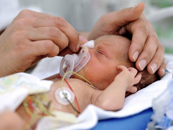
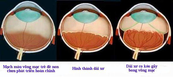
During the last 12 weeks of pregnancy, eyes develop rapidly. If the baby is born at term, the development of the retinal blood vessels is almost complete (the retina usually ends a few weeks to a month after birth). But if a baby is born early, before these blood vessels grow to the edge of the retina, normal growth can stop. As a result, the periphery may not receive enough oxygen and nutrients. As a result, abnormal circuits begin to develop. These abnormal new blood vessels are fragile and vulnerable to risk factors (such as oxygen poisoning) that cause bleeding and scarring in the retina. As this scar shrinks, they pull on the retina causing it to separate from the bottom of the eye (retinal detachment). Therefore, children need to be screened for retinopathy of prematurity to limit complications.
This disorder usually develops in both eyes and is one of the common causes of vision impairment or permanent blindness.
2. Time to check Rop for premature babies
Follow up every 1-2 weeks Depending on the progression of the disease, follow up until the retina matures, the disease completely regresses or there is an indication for treatment.
3. Stages of retinopathy of prematurity
Stage 1: mild vascular growth. A thin white line appears, separating the two areas: the vascularized retina and the avascular retina. In stage 1, the blood vessels can continue to develop normally, but the doctor needs to closely monitor the child's condition. Many children with stage 1 disease improve without treatment and eventually develop normal vision. The disease resolves on its own with no further progress Stage 2: Moderately abnormal blood vessel growth. At this time, the boundary line between the vascularized retina and the avascular retinal area became more visible and grew away from the retinal surface, becoming wide and high forming a white ridge ( if few blood vessels) or pink (if many blood vessels). Many children who develop at this stage are also able to improve on their own without treatment.
Stage 3: Severe abnormal blood vessel growth. This is the stage of extraretinal vascular fibrillation. From the surface of the ridge, fibrovascular tissue proliferates and spreads to the posterior surface of the retina; or develop anteriorly, perpendicular to the retinal plane into the vitreous. In stage 3, ROP disease is also divided by mild, moderate and severe; depending on the extent of proliferation of vascular tissue into the vitreous.
* Stage 4B: eye function is markedly reduced. The retinal detachment is more extensive and extends to the retina in the macular region.
Stage 5: Total retinal detachment. The detached retina has a funnel shape. There are three types of hopper: open hopper, closed hopper, front closed, rear open. Possibility of permanent blindness.
4. Treatment of retinopathy of prematurity
* AVATIN is a class of angiogenesis inhibitor monoclonal antibody A (VEGF-A). has been used by many countries in the world such as the US, Canada, Mexico, Chile, Germany, the Netherlands, Turkey, Taiwan, Thailand... and Vietnam in the treatment of retinopathy of prematurity and for very good results. Thanks to this treatment method, thousands of children around the world can avoid blindness due to ROP. Intraocular injection of Avastin. The drug can be used early before laser treatment or used late when laser treatment fails. Usually the best results are at stage 3+
*Laser therapy burns away the peripheral areas of the retina that do not have normal blood vessels. This treatment destroys the peripheral area of the retina, slowing or reversing the abnormal growth of blood vessels.
For children with more severe disease, other treatments are performed but the results are not satisfactory.
Removal of the vitreous in the eye: This involves removing the vitreous or replacing it with saline. After the vitreous is removed, the scar tissue on the retina can be peeled off or cut away, allowing the retina to no longer stretch.
Membrane flexion: This method will use a silicone band around the eye and tighten it will prevent the vitreous gel from pulling on the scar tissue and allow the retina to flatten
Retinopathy of prematurity is treatable and If the disease is treated early, it is highly effective, so premature babies need to be screened for regular eye exams under the guidance of a doctor.

Parents should not breastfeed their babies 1 hour before the eye exam to avoid vomiting and inhaling dangerous foods.
To accurately examine the baby's eyes, the baby needs to drop copper dilating medicine 3-4 times, so it takes 45-60 minutes for the medicine to take effect. Therefore, parents should choose the right time to visit the hospital.
At Vinmec International Hospital, we are applying laser treatment of retinal detachment and Avastin injection with the advantage of improving visual function for children. patient.
Để đặt lịch khám tại viện, Quý khách vui lòng bấm số HOTLINE hoặc đặt lịch trực tiếp TẠI ĐÂY. Tải và đặt lịch khám tự động trên ứng dụng MyVinmec để quản lý, theo dõi lịch và đặt hẹn mọi lúc mọi nơi ngay trên ứng dụng.
Dịch vụ từ Vinmec
-
 Tại sao bạn nên xử trí viêm màng bồ đào sớm?
Viêm màng bồ đào có thể dẫn đến các vấn đề nghiêm trọng về mắt nếu không điều trị ngay lập tức. Đặc biệt nếu mắc bệnh trong một thời gian dài, hoặc mắc bệnh khi ở tuổi già thì ...
Đọc thêm -
 Điều trị hạch mạc treo ở trẻ 4,5 tháng tuổi bị trào ngược dạ dày?
Con trai em được 4,5 tháng, bú sữa ngoài, bị nôn trớ, trào ngược dạ dày từ khi mới sinh. Bé thường xuyên bị nhiều hơi trong ruột. Hơn 1 tuần nay bé không chịu bú sữa, ép uống thì ...
Đọc thêm -
 Trẻ sơ sinh bị viêm ruột kèm phân có dịch nhầy, trắng là triệu chứng bệnh gì?
Bé nhà em 4 tháng đi ngoài phân nhầy và máu kèm sốt cao, gia đình đưa cháu đi khám và điều trị tại bệnh viện khoảng 1 tuần và bác sĩ kết luận bé bị viêm ruột. Sau khi ...
Đọc thêm -
 Trẻ sinh non tính theo tháng tuổi nào để tiêm chủng?
Con em sinh non lúc 27 tuần 2 ngày. Bé sinh ngày 12/8/2019, con trai ạ. Bé chỉ được tiêm phòng lao. Từ đó tới nay chưa tiêm mũi nào nữa ạ. Bé sinh non có tiền sử viêm phổi ...
Đọc thêm -
 Bong võng mạc do sinh non chữa được không?
Mắt cháu bị bong võng mạc do sinh non, một mắt không nhìn thấy còn một mắt cận nặng. Hai mắt đều bị teo võng mạc. Bác sĩ cho cháu hỏi bong võng mạc do sinh non chữa được không?
Đọc thêm