
Understanding the stages and types of breast cancer
Breast cancer is the most common and leading cause of death in women. In particular, staging is a very important prognostic factor in breast cancer treatment. To be able to accurately assess the stage, doctors need to do a physical examination and use a variety of imaging tests to survey the structure and extent of invasion of the tumor.
1. What is breast cancer?
Breast cancer is cancer that forms in the cells of the breast. After skin cancer, breast cancer is the most common cancer diagnosed in women in the United States. Breast cancer can occur in both men and women, but it is more common in women. Breast cancer is closely related to genetic mutations (such as BRCA1 and BRCA2), the actions of female sex hormones such as estrogen and progesterone.
Substantial support for breast cancer awareness and research funding has helped create advances in breast cancer diagnosis and treatment. Breast cancer survival rates have increased and the number of deaths related to the disease is decreasing, largely due to factors such as earlier detection, new personalized treatments, and understanding. know better about this disease.
In which, the assessment of breast cancer stage is one of the very important factors to choose the appropriate breast cancer treatment method. Breast cancer stage describes how far the cancer has spread in the breast and other parts of the body.

2. What is the staging of breast cancer?
Staging breast cancer is based on tumor size and how far the cancer has spread to other parts of the body. Your doctor will diagnose breast cancer after doing a physical exam and using a number of imaging tests (such as mammogram, breast ultrasound, CT-scan, MRI, or PET-CT) to help diagnose your breast cancer. assess the extent of breast cancer.
After complete clinical and laboratory investigations, the doctor will make a staging diagnosis. If staging is operable, your doctor will likely stage your breast cancer again after surgical removal of the tumor to determine the exact stage of the cancer after surgery. This is a very important step in assessing whether other adjuvant treatments such as chemotherapy or radiation are needed.
The simplest approach to interpreting breast cancer staging is to use the T, N, and M classifications. This is the approach used below to describe the different stages.
Most patients are anxious to find out the exact stage of the cancer. If the patient has surgery as the first treatment, the doctor will confirm the cancer stage after surgery. Tissue samples are taken and tested, results will be answered usually about 5 to 7 days after surgery.
3. TNM classification system for breast cancer
To be able to unify staging in cancer, major associations around the world have agreed to use the TNM classification system to assess the invasiveness of cancer.
Through clinical examination and imaging tests, the doctor will evaluate the three elements of this classification system including tumor (Tumor), lymph nodes (Node) and distant metastasis (Metastasis) .
Tumor (T): size and location of tumor Node (N): Has there been lymph node metastasis? Size, number and location of metastatic nodes Metastasis (M): whether the cancer has spread to other organs of the body The TNM classification of breast cancer is as follows:
T: Primary tumor: Tis: Pre-invasive epithelial tumor or Paget's disease of the nipple.
T0: No palpable breast tumor
T1: Tumor is ≤ 2 cm in greatest size including T1a, T1b
T1a U is not attached to the fascia or pectoralis major
T1b U is attached to the fascia or pectoralis major
T2: U with size > 2 cm, ≤ 5 cm including T2a, T2b
T3: Tumor with tumor size > 5 cm including T3a, T3b
T4: Tumor spreads directly into the chest wall or into the skin regardless of size. Includes T4a, T4b, T4c
N: Regional lymph nodes: Nx: Clinically unidentified lymph nodes
N0: No palpable axillary lymph nodes
N1: ipsilateral axillary lymph nodes are mobile
N2: ipsilateral ipsilateral axillary lymph nodes into or into another organ
N3: Lymphadenopathy supraclavicular or subclavian hand edema, and/or ipsilateral internal mammary nodes.
M: Distant metastasis: M0: No distant metastasis
M1: Distant metastasis including spread beyond the breast

4. Histological grading of breast cancer
The grading system varies depending on the type of cancer. In general, tumors are graded as 1, 2, 3, or 4, depending on the extent of the abnormality. Grade 1 tumors have cancerous cells and tissues that appear nearly normal (well differentiated). These tumors tend to grow and spread slowly. In contrast, grade 3 and 4 cancer cells and tissues have a shape and structure unlike normal cells and tissues, and they tend to grow and spread more quickly.
If the histopathological grade of the tumor is not specified separately, the following system is commonly used (1):
GX: Unknown (Type Unknown) G1: Well differentiated (low grade) G2: Moderately differentiated (moderate) G3: Poorly differentiated (high level) G4: Undifferentiated (high grade) For breast cancer, doctors usually use the Nottingham grading system (also known as the Nottingham grading system). is an Elston-Ellis modification of the Scarff-Bloom-Richardson grading system) for breast cancer patients. This system classifies tumors based on the following characteristics:
Tubularity: what percentage of cancer tissue forms ductal structures Cell nucleus: assesses the size and shape of nuclei in cells cancer Mitotic rate: how many cells divide, this is a measure of how quickly cancer cells grow and divide Each item is scored from 1 to 3 points; 1 point means cancer tissues and cells are the same as normal tissues and cells; 3 points mean the most abnormal cells and tissues. Then add up and total score from 3 to 9:
Total score = 3-5: G1 (low or well differentiated) Total score = 6-7: G2 (moderate or moderately differentiated) Total score = 8-9: G3 (high level or poorly differentiated)
5. Staging of breast cancer
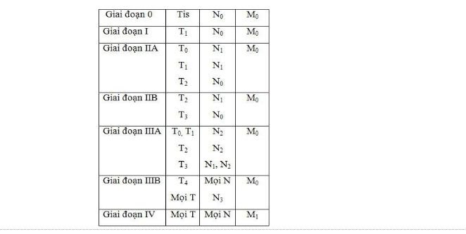
After answering the above three factors, the stage of breast cancer will be graded based on the level of these three factors. For breast cancer, there are 5 stages including stage 0 (zero) which is non-invasive ductal carcinoma in situ (DCIS) and stages I to IV (1 to 4), which is used for breast cancer. invasive breast.
Staging can be clinical or pathological (postoperative). Clinical staging is based on the results of tests performed prior to surgery, which may include a physical exam, mammogram, ultrasound, and MRI scan. Pathology segmentation is based on what is found during the surgical removal of breast tissue and lymph nodes. Results are usually available a few days after surgery. In general, the pathological (postoperative) staging provides the most information for determining a patient's prognosis.
Doctors will determine the stage of cancer by combining T, N and M classifications, histological grade and test results of tumor biomarkers (estrogen receptor - ER / progesterone receptor). - PR and HER2. This information is used to help determine the prognosis of breast cancer Breast cancer staging includes:
Stage 0 refers to 'pre-invasive' breast cancer, which includes breast cancer. ductal carcinoma in situ (DCIS) Stages I and II are called early breast cancer Stage III is called locally advanced breast cancer Stage IV is called breast cancer advanced or metastatic. At this stage, the cancer has spread to other parts of the body. Detailed classification of breast cancer stages.

6. Breast cancer prognosis
Prognosis refers to the probable or likely outcome of a disease (or chance of recovery). While tumor stage and grade are important, doctors will also take other considerations into account, such as whether certain molecular receptors are present, when determining a patient's prognosis for breast cancer.
While it is impossible to predict the exact course of the disease for any individual, survival rates for breast cancer have improved dramatically over time due to earlier detection and treatments. improved. The five-year survival rate for breast cancer is currently 91%. Most patients with early-stage or late-stage breast cancer in situ (Stage 0 - 3) can be successfully treated.
At Vinmec International General Hospital, there is a Breast Cancer Screening Package, which helps customers screen and detect breast cancer early even when there are no symptoms. When registering for the Breast Cancer Screening Package, customers will receive:
Examination and consultation with an oncologist. Breast cancer screening by bilateral breast ultrasound and mammogram. Officially put into operation on January 7, 2012, Vinmec has become a prestigious address in breast cancer screening with:
Team of highly qualified and experienced doctors. Comprehensive professional cooperation with domestic and international hospitals: Singapore, Japan, USA, .. Comprehensive treatment and care, multi-specialty coordination towards individualizing each patient. Having a full range of specialized facilities for diagnosis and staging before treatment: Endoscopy, CT scan, PET-CT scan, MRI, histopathological diagnosis, gene-cell testing, .. There are a full range of mainstream cancer treatment methods: surgery, radiation therapy, chemotherapy, stem cell transplant...
Để đặt lịch khám tại viện, Quý khách vui lòng bấm số HOTLINE hoặc đặt lịch trực tiếp TẠI ĐÂY. Tải và đặt lịch khám tự động trên ứng dụng MyVinmec để quản lý, theo dõi lịch và đặt hẹn mọi lúc mọi nơi ngay trên ứng dụng.
Dịch vụ từ Vinmec
-
 Ung thư gây ra các biến chứng đe dọa tính mạng như thế nào?
Ung thư là một căn bệnh xảy ra khi những tế bào bất thường phát triển không kiểm soát được. Vậy ung thư sẽ gây nên các biến chứng đe dọa tính mạng như thế nào? Bị ung thư có ...
Đọc thêm -
 Ung thư giáp di căn hạch uống Iod 131 hiệu quả không?
Em bị ung thư giáp đã mổ gần được 2 tháng, nay xét nghiệm và siêu âm sau 3 tuần kiêng Hocmon thấy bị suy giáp (TSH <100) và có vài hạch nhóm IV nghi di căn. Bác sĩ hội ...
Đọc thêm -
 Những điều cần biết về ung thư đại trực tràng di truyền
Đột biến di truyền làm tăng nguy cơ mắc một hoặc nhiều loại ung thư của một cá thể và được truyền từ bố mẹ sang con cái. APC, EPCAM, MLH1, MSH2 và MSH6 là những gen làm tăng nguy ...
Đọc thêm -
 Bạn có nên thực hiện xét nghiệm di truyền để đánh giá nguy cơ ung thư?
Khoảng 5 – 10% các trường hợp ung thư được cho là có liên quan tới các đột biến gen có khả năng di truyền hoặc truyền lại trong gia đình. Việc mang các đột biến có tính chất di ...
Đọc thêm -
 Các biểu hiện ung thư thực quản có dễ nhận biết?
Ung thư thực quản là một trong những căn bệnh ung thư thường gặp ở đường tiêu hóa. Đối với những biểu hiện bệnh khác nhau trong từng giai đoạn, ung thư thực quản thường rất khó phát hiện trong ...
Đọc thêm