
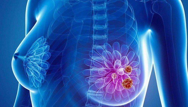
How is breast cancer diagnosed?
1. What is breast chlorophyll?
It is estimated that phyllodes tumors account for less than 1% of all breast tumors. Of which, 90% of chlorophyll tumors in the mammary gland are not malignant tumors, the remaining 10% have the potential to develop into breast cancer. However, no matter what type, syphilis in the breast tends to grow very quickly and all require treatment.
Currently, the cause of the formation of chlorophyll tumors in the breast is still not clear, only known that chlorophyll in the breast can appear at any age, but it is most common in women above 40 years old and not yet menopause. In addition, there are factors that can increase the risk of disease such as: people who are breastfeeding, people who have physical damage to the chest area.

2. Symptoms of breast chlorophyll tumor
The average tumor size is 4-7cm, the smallest tumor is only 1cm. but can be as large as 40cm, usually appearing in the upper quadrant of the udder. Breast phyllodes tumor has a rapid growth rate, increasing 2-3cm in a few months. When palpating the phyllodes tumor, it will be firm, clear, well mobile even though the tumor is large, multi-lobed and painless, the skin on the tumor is usually smooth, glossy and thin. There are rare cases of chest wall invasion, nipple retraction, skin ulceration, and rarely bilateral tumors.
3. Diagnosis of breast chlorophyll tumor

The edges of the tumor are clearly defined. Cells do not tend to multiply too quickly. Connective tissue cells look like normal cells and do not overgrow. * Malignant chlorophyll tumors (about 25%) will have the following characteristics:
Tumor has undefined edges. Cells that divide rapidly, have irregular shapes. Connective tissue cells overgrow. Breast chlorophyll tumors can be located between benign and malignant, and will have mixed features of both types.
3.2. Methods of diagnosis of breast chlorophyll tumors Currently, to diagnose breast chlorophyll tumors, common diagnostic methods are applied such as:
Paraclinical examination: palpation to assess the abnormality, examination Check the shape and size of the breast. Breast ultrasound: helps to observe the nature and shape of the tumor. Mammography (Mammography): Breast phyllodes tumor usually appears as a large round or oval shape with clear lobe margins. Mammography can be combined with other tests to confirm the correct diagnosis. Mammography MRI: helps to see more clearly the image of the tumor and plan the surgical route. Breast biopsies include 2 types: Core biopsy - take a sample of the tumor and aspirate to remove the entire tumor after being identified by Vacuum Needle and testing for tumor cells.

At Vinmec International General Hospital, there is a Breast Cancer Screening Package, which helps customers screen and detect breast cancer early even when there are no symptoms. When registering for the Breast Cancer Screening Package, customers will receive:
Examination and consultation with an oncologist. Breast cancer screening by bilateral breast ultrasound and Mammography (mammogram). Up to now, Vinmec has become a prestigious address in breast cancer screening with:
Team of highly qualified and experienced doctors. Comprehensive professional cooperation with domestic and international hospitals: Singapore, Japan, USA, etc. Comprehensive treatment and care for patients, multi-specialty coordination towards individualizing each patient. Having a full range of specialized facilities to diagnose the disease and stage it before treatment: Endoscopy, CT scan, PET-CT scan, MRI, histopathological diagnosis, gene-cell testing, .. There are full range of mainstream cancer treatment methods: surgery, radiation therapy, chemotherapy, stem cell transplant...
Để đặt lịch khám tại viện, Quý khách vui lòng bấm số HOTLINE hoặc đặt lịch trực tiếp TẠI ĐÂY. Tải và đặt lịch khám tự động trên ứng dụng MyVinmec để quản lý, theo dõi lịch và đặt hẹn mọi lúc mọi nơi ngay trên ứng dụng.
Dịch vụ từ Vinmec
-
 Vú xuất hiện khối u và thay đổi hình dạng có đáng lo?
Cho cháu hỏi là vú của cháu có 1 cục hơi to và vú có hình dạng hơi xẹp xuống, đó có phải là triệu chứng gì không ạ?
Đọc thêm -
 Thời điểm thích hợp chụp cộng hưởng từ tuyến vú
Chụp cộng hưởng từ tuyến vú là phương pháp chẩn đoán hình ảnh hiện đại có sử dụng từ trường, sóng vô tuyến và máy tính để tạo ra các hình ảnh chi tiết về tuyến vú. Đây là kỹ ...
Đọc thêm -
 Sau sinh mổ xuất hiện u trên ngực có nguy hiểm không?
Em mới sinh mổ được 20 ngày, cách đây gần nửa tháng em phát hiện bên ngực trái phía trên có hạch, sờ thấy khác biệt hẳn so với bên ngực phải. Bác sĩ cho em hỏi dấu hiệu này ...
Đọc thêm -
 Phụ nữ cho con bú có thể sàng lọc ung thư vú không?
Vợ em bị đau nhức một bên vú, đi siêu âm thì phát hiện khoảng giảm âm 5x9mm. Vợ em đang cho con bú 7 tháng. Vậy bác sĩ cho em hỏi phụ nữ cho con bú có thể sàng ...
Đọc thêm -
 Điều trị ung thư vú giai đoạn 4, bướu độc liệu mô như nào?
Bác sĩ cho em hỏi phát hiện ung thư vú giai đoạn 4, bướu độc liệu mô thì tỷ lệ khỏi như thế nào? Phương pháp điều trị ung thư vú giai đoạn 4, bướu độc liệu mô như nào?
Đọc thêm