
Imaging features and value of multislice computed tomography in chronic pancreatitis
Posted by Master, Doctor Pham Manh Chung - Head of Imaging Department - Department of Diagnostic Imaging - Vinmec Ha Long International Hospital
Chronic pancreatitis is a pathological process with a long continuum, the pancreas is damaged leading to destruction of the pancreatic parenchyma or pancreatic duct and replaced by fibrous tissue leading to impaired pancreatic function. The diagnosis of chronic pancreatitis based on the imaging features of the multi-slice computed tomography machine will help doctors identify inflammation as well as lesions in the pancreas for effective treatment.
1. What is chronic pancreatitis?
Chronic pancreatitis (VTM) is an inflammatory disease characterized by progressive, irreversible destruction of the pancreatic parenchyma, gradually leading to fibrosis of the pancreas with clinical manifestations of chronic abdominal pain, emaciation, and emaciation. weight loss and decreased endocrine and exocrine pancreatic function.
2. Symptoms and manifestations of chronic pancreatitis
Chronic pancreatitis has very clear symptoms, specifically as follows:
Abdominal pain: Abdominal pain is the main clinical symptom in VTE, encountered in most patients, which is also the main reason why patient admitted to the hospital. The disease manifests as abdominal pain above the navel, spreading to the back, more painful after eating, can be persistent and difficult to treat. The disease can cause pain in episodes with variable intensity, sometimes the pain is repeated many times, sometimes the dominant pain is described as a sharp pain, the patient has to change position to pain relief “VTM pain relief posture” Weight loss: Weight loss in the early stages is because the patient is afraid to eat because it is related to pain. In the later stage, it is due to malabsorption because of the lack of exocrine pancreatic enzymes to break down proteins and lipids. Poor eating and anorexia lead to exhaustion, most patients with chronic pancreatitis are thin because they have malabsorption. Digestive disorders: Prolonged diarrhea or greasy, raw stools cause malabsorption syndrome. Jaundice: Due to narrowing of the common bile duct leading to biliary obstruction, jaundice may be intermittent or progressive, jaundice often accompanied by pain may also be transient.

Diabetes: The rate of chronic pancreatitis with diabetes accounts for about 30%. Diabetes mellitus in chronic pancreatitis is caused by impaired secretion of the hormone insulin involved in glucose metabolism in the body.
3. Causes of chronic pancreatitis
There are many causes of chronic pancreatitis, specifically:
3.1 Alcoholic chronic pancreatitis
Alcohol is responsible for 70-80% of cases of chronic pancreatitis. There is no threshold for the toxic effects of alcohol on the pancreas, but the amount and duration of alcohol consumption correlates with the development of chronic pancreatitis. Some studies suggest that taking 150-200ml daily for 10 to 15 years is required for the development of chronic pancreatitis. Alcohol-related causes are common in Europe, America, Central Africa and also in Asia, mainly in men, these patients with chronic pancreatitis have an average age of 36-55 years.

3.2. Idiopathic chronic pancreatitis
Idiopathic chronic pancreatitis accounts for about 20-30% of patients with idiopathic pancreatitis, this number is referred to as primary chronic pancreatitis. Epidemiological evidence suggests that chronic idiopathic pancreatitis is a distinct form, equally affecting both men and women, and that slow progression of endocrine and exocrine pancreatic insufficiency has been observed.
Some other causes of chronic pancreatitis include:
Chronic tropical pancreatitis. Increased blood calcium. Increased blood lipids. Inherited chronic pancreatitis: caused by mutations in the trypsinogen gene, mutations or changes in the trypsin secretion inhibitor gene, the cystic fibrosis gene. Gallstones: VTE is associated with gallstones, worms in the bile ducts, cholecystitis, stricture of the ampulla of Vante, and gallstones of the ampulla of Vante. This cause is common in Asian countries such as China and Vietnam. Chronic pancreatitis due to pancreatic tumors. Autoimmune chronic pancreatitis

Causes of pancreatic stones: there may be stones in the pancreatic duct or calcification of the pancreatic parenchyma in the head, body or tail or calcification throughout the pancreas. Causes of acute pancreatitis : Severe acute pancreatitis recurs gradually leading to pancreatic fibrosis and calcium deposition of pancreatic parenchyma causing chronic pancreatitis.
4. Imaging characteristics and value of multislice computed tomography in the diagnosis of chronic pancreatitis.
Multi-slice computed tomography is an increasingly used technique in the diagnosis of pancreatic diseases due to advances in technology, imaging speed and image resolution. Multi-slice computed tomography scans well assess the pancreatic parenchyma, size, border, pancreatic duct and affect the peripancreatic organs such as biliary tract, duodenum, peripancreatic spaces.
4.1. Signs of chronic pancreatitis on computed tomography
Change in size of pancreas: In the early stages of VTM, the size of the pancreas may be normal. In advanced stages, the pancreas is often diffuse or focal. In the late stage, most of the people see pancreatic atrophy. Pancreatic border: irregular due to fibrosis and calcification of the pancreatic parenchyma, infiltrating the fatty layer around the pancreas. Pancreatic stones: Computed tomography is the best measure to identify pancreatic stones. Stones larger than 1mm can be seen on CT scan, stones appear in the parenchyma or in the pancreatic duct or in both the pancreatic parenchyma and the pancreatic duct. Dilatation of the main pancreatic duct: the pancreatic duct is visible when the diameter is > 3 mm on radiographs with thin sections. The characteristic feature of pancreatic ductal dilatation in VTM is irregular dilation, with many narrow segments interspersed with dilated segments, creating a rosary-like image.

Pancreatic pseudocyst: manifested by low density structures (0-20HU), well-defined boundaries, located in pancreatic parenchyma or extrapancreatic sites, no enhancement after injection. If the pancreatic pseudocyst is complicated by intracystic bleeding or the cyst is superinfected, the density in the cyst may be higher. Narrowing of the main intrapancreatic bile duct: when the inflammatory process is concentrated in the head of the pancreas, the fibrous tissue proliferates, compressing and narrowing the intrapancreatic bile duct, and causing dilation of the upper bile duct at the narrowing site. Duodenal stenosis: due to compression of pancreatic head by fibrosis or pancreatic pseudocyst. Manifested by change in diameter of the duodenum and dilatation of the stomach and duodenum above the stricture. Duodenal stenosis is often associated with biliary dilatation. Vascular lesions: thrombosis of the splenic vein, superior mesenteric vein, portal vein thrombosis, often associated with signs of portal hypertension. Pseudoaneurysm is common in splenic artery pseudoaneurysm, the manifestation of pseudoaneurysm mass is the density of blood vessels in the arterial phase. VTE exacerbation: manifestations on CT scan are increased pancreatic size, peripancreatic fat infiltration, pararenal fascia thickening, fluid accumulation in the omentum omentum, anterior pararenal space, perirenal space, mesenteric roots, mesenteric mesentery transverse colon. Pancreatic outflow follows the left pararenal space down to the pelvis, and the mesenteric roots of the small intestine.

4.2. Value of multislice computed tomography in the diagnosis of chronic pancreatitis
This is a non-invasive imaging technique that can cut thin layers of less than 1mm, has the ability to reconstruct in many planes, allowing accurate assessment of lesions in the pancreas, biliary system, and system. vascular system and surrounding organs to make an accurate diagnosis of chronic pancreatitis, and based on computed tomography images, clinicians will provide accurate and appropriate treatment.
5. Why should multi-slice computed tomography at Vinmec International General Hospital?
Before multi-sequential computed tomography, you will be evaluated by leading medical experts on your medical history, personal history, safety check for computed tomography and contrast injection (check list of CT scans). ) to ensure the safety of the client before entering the tomography room and injecting contrast.
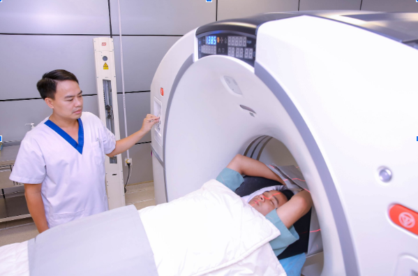
The computerized tomography procedure performed by a team of good and professional doctors and technicians will make you confident in your expertise, peace of mind and comfort before, during and after the scan. Especially now Vinmec is equipped with a 640-slice CT scanner TSX-301C manufactured by Toshiba capable of supporting diagnosis on an area up to 16cm wide, the machine will cut slices as thin as 0.5mm for full evaluation. hurt. With high quality machine will greatly reduce the radiation dose for customers.
Để đặt lịch khám tại viện, Quý khách vui lòng bấm số HOTLINE hoặc đặt lịch trực tiếp TẠI ĐÂY. Tải và đặt lịch khám tự động trên ứng dụng MyVinmec để quản lý, theo dõi lịch và đặt hẹn mọi lúc mọi nơi ngay trên ứng dụng.
Dịch vụ từ Vinmec
-
 Trẻ 4 tuổi rưỡi bị viêm tụy cấp đã kiêng dầu mỡ và các thức ăn nhiều protein nhưng vẫn bị đau bụng là do đâu
Chào bác sĩ! Con cháu năm nay 4,5 tuổi, đi khám chẩn đoán bị viêm tụy cấp, giờ bé chỉ ăn cháo đường, cháo muối, nhưng 2 tuần nay rồi bé vẫn thỉnh thoảng đau bụng, mỗi lần đau khoảng ...
Đọc thêm -
 Nữ giới đau vùng thượng vị, chướng bụng, buồn nôn là dấu hiệu bệnh gì?
Em bị đau ngay thượng vị, chướng bụng, khó thở, tức ngực, buồn nôn, đau nhiều về đêm. Em đi khám thì có kết quả bị xung huyết niêm mạc hang vị (mức độ nhẹ), em vẫn uống thuốc điều ...
Đọc thêm -
 Sau điều trị viêm tụy mạn bị sốt có nên đến bệnh viện không?
Chào Bác sĩ! Bác sĩ cho em hỏi, bố em bị viêm tụy mạn đã điều trị ở 2 bệnh viện lớn, tạm ổn rồi về nhà được 5 ngày thì bị sốt. Bố em có bệnh lý kèm là ...
Đọc thêm -
 Viêm tụy mạn là gì? Có nguy hiểm không?
Có câu hỏi này mong bác sĩ trả lời sớm để em có hướng điều trị kịp thời. Ngày 10/3/2020 em có bị nặng bụng phía trên. Em đi khám 2 lần. Lần đầu tiên em nội soi bao tử ...
Đọc thêm -
Viêm tụy mạn là gì? Có nguy hiểm không?
Có câu hỏi này mong bác sĩ trả lời sớm để em có hướng điều trị kịp thời. Ngày 10/3/2020 em có bị nặng bụng phía trên. Em đi khám 2 lần. Lần đầu tiên em nội soi bao tử ...
Đọc thêm