
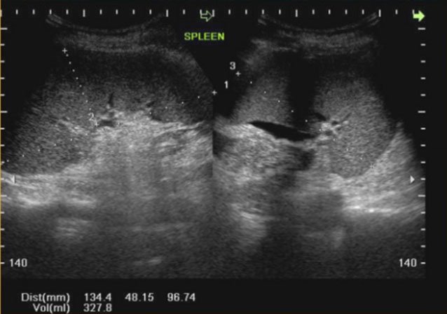
Ultrasound image of splenomegaly
The article is professionally consulted by Master, Doctor Lam Thi Kim Chi - Department of Diagnostic Imaging - Vinmec Danang International General Hospital.
The spleen is an organ of the body, hidden in the abdomen, just below the ribcage, normally not palpable except in the case of young children, the abdominal wall is flat. On clinical examination, only the opaque area of the spleen can be identified in the posterior axillary line. In case the spleen is enlarged, it crosses the costal margin, the doctor can feel it. Today, thanks to the development of science and technology, ultrasound has helped diagnose more accurately.
1. What is an enlarged spleen?
The spleen is a small organ located deep in the abdomen, hiding under the left diaphragm. Normally we cannot feel the spleen, except in the case of young children, the abdominal wall is flat. The spleen is involved in the production of lymphocytes to protect the body, in addition, at the fetal stage, the spleen also produces granulocytes, red blood cells and platelets.
The spleen is the site of destruction of old blood cells, retaining iron, protein and other substances needed to make new blood cells. This is also where the body stores blood. When the spleen contracts or dilates, it participates in the regulation of blood volume as well as the volume of blood cells in the circulation. In addition, the spleen also participates in fighting infections for the body by purifying bacteria and foreign objects in the blood.

Therefore, the spleen can be enlarged in many different diseases. With only one symptom of splenomegaly, it is not possible to diagnose the cause of the disease. However, when the spleen is larger than normal, it means it must have been overactive.
2. Diagnosis of splenomegaly on ultrasound
Ultrasound of the spleen is difficult because the spleen is smaller than the liver, it is located close to the diaphragm and is covered by the ribs. However, through the intercostal space, the ultrasound image also shows the host splenic tissue and measures its size. Although the spleen is well evaluated by computed tomography and magnetic resonance imaging; Splenic ultrasound has advantages such as ease of implementation, non-invasiveness, low cost, and can be performed at the bedside in case of emergency.
Indications for splenic ultrasound in the following cases:
Verify splenomegaly and monitor the progression of the spleen through treatment Detecting splenomegaly with uniform echo, diffuse or regional echogenic changes, thereby impaired disease of the spleen. Detection of hematoma or splenic rupture after trauma. Cases of unexplained pain in the left upper quadrant of the abdomen. Prepare the patient
Fasting for at least 6 hours Do not use Baryte for gastrointestinal imaging Do not perform laparoscopy before ultrasound. Practice for the patient to exhale and hold his breath during the ultrasound. When necessary, the patient can be given water to drink to separate the spleen from the left lobe of the liver. A normal ultrasound image will show the spleen as truncated semi-conical, concave on the medial surface, convex on the outer surface, with the following dimensions:
Length (L) ≤ 12cm in adults. For children from 0 to 3 years old, the length of the spleen is less than 6cm. For children, the length of the spleen is calculated as follows: L = 5.7 + 0.31 x age (in years). Thickness (T) ≤ 7cm. Width (W) ≤ 5cm. Spleen index = L x T x W < 480 Spleen weight (SW) = Spleen index x 0.55. The spleen at birth is about 15g, the adult spleen is about 150g, ranging from 100-265g. However, the determination of splenomegaly on ultrasound still has many different opinions, some authors agree with the above size, that is 12cm (length) is the maximum size. However, there are other authors who believe that the length of the spleen can be up to 14cm.
Here, you can refer to the assessment of splenomegaly based on spleen weight as follows:
Splenomegaly: SW <500g Moderate spleen: 500

3. Causes of splenomegaly
Because the spleen has many different tasks, when detecting a patient with splenomegaly, the doctor needs to ask, do a thorough clinical examination and maybe do some more tests towards the pathology of the blood and muscles. other hematopoietic organs, diseases of the liver and portal vein circulation. Because the causes of splenomegaly are many, the most important are still two main groups of causes: blood diseases and diseases of the liver and portal vein system.
Usually splenomegaly is divided into two types:
Chronic splenomegaly. Acute splenomegaly.
3.1. Chronic Splenomegaly This is the most common type of splenomegaly, and the cause is more difficult to diagnose.
Enlarged spleen in blood diseases:
Neoplastic leukemia. Hodgkin's disease. Besnier-Boeck Schaumann disease. Disseminated sarcoma: Splenomegaly with erythrocytosis: Vaquez disease. Spleen tuberculosis. Splenomegaly with increased myelocytosis: medullary crystalline leukocytes.
Enlarged spleen with hemolytic anemia syndrome
Congenital hemolytic anemia (Minkowski - Chauffard disease). Hemolytic anemia due to blood diseases of hemoglobin: Hemolytic anemia with target-shaped red blood cells, marine anemia. Sickle cell hemolytic anemia. Acquired hemolytic anemia: caused by toxicity, infection, parasites and malignancies. Splenomegaly with decreased blood cells: this is a syndrome of primary or secondary total cell anemia. Splenomegaly due to liver disease, portal vein. Splenomegaly due to other causes, often difficult to diagnose, such as:
Parasitic infections. Chronic fever. Hidden Leukemia. Banti disease (primary splenomegaly) in the early stages. Chronic bacterial infections such as tuberculosis. Splenic cysts, malignant or benign tumors. Fat disorders cause stagnation in the spleen.

3.2. Acute splenomegaly Acute splenomegaly is splenomegaly that is only an immediate sign or a secondary sign in a multi-characteristic setting.
Blood infection caused by common pus bacteria. Typhoid or paratyphoid disease. Acute or subacute endocarditis, tuberculosis, rickettsial spirochetes There are three acute diseases in which splenomegaly is an important symptom, which are diseases:
Acute leukemia . Osler's endocarditis. Infectious mononucleosis. It can be seen that splenomegaly is a common clinical sign, the diagnosis of splenomegaly on ultrasound is usually easy, but to diagnose the cause is often very difficult. However, for a patient with splenomegaly, it is necessary to think a lot about blood diseases and liver diseases, especially cirrhosis. To find the cause, it is often necessary to have a careful examination combined with some other necessary tests.
MSc Doctor Lam Thi Kim Chi graduated with a Master's degree in Radiology - Hue University of Medicine and Pharmacy and has over 6 years of experience as a radiologist. Doctor Chi used to work at Da Nang Obstetrics and Gynecology Hospital before working as a radiologist at Vinmec Danang International General Hospital as it is today.
Customers can directly go to Vinmec Health system nationwide to visit or contact the hotline here for support.
Dịch vụ từ Vinmec
-
 Hậu quả của cường lách do xơ gan
Xơ gan là một căn bệnh có tiến triển một cách âm thầm nhưng gây ra những biến chứng vô cùng nguy hiểm. Cường lách do xơ gan chính là một trong những biến chứng nguy hiểm do xơ gan ...
Đọc thêm -
Cường lách gây ảnh hưởng gì?
Lá lách là cơ quan nằm ngay dưới lồng xương sườn bên trái của bạn. Có nhiều nguyên nhân như nhiễm trùng, bệnh gan và một số bệnh ung thư có thể gây ra lách to hay còn gọi là ...
Đọc thêm -
Chỉ định của phẫu thuật nội soi cắt lách
Với các trường hợp có vấn đề bất thường vùng lách, cần kiểm tra, chẩn đoán và điều trị sớm để tránh nguy cơ tai nạn vỡ lách gây nguy hiểm tới tính mạng. Mổ nội soi cắt lách là ...
Đọc thêm -
Xơ gan lách to kiểu Banti là gì?
Xơ gan lách to kiểu Banti là một bệnh lý của xơ gan, gây lách to và giảm số lượng tế bào máu. Nếu phẫu thuật cắt lách khi gan chưa xơ hoặc xơ nhẹ sẽ ngăn chặn được sự ...
Đọc thêm -
 Các bước thực hiện siêu âm chẩn đoán bệnh lý lách
Có rất nhiều bệnh lý về lá lách có thể được xác định thông qua quá trình siêu âm. Quá trình siêu âm lá lách diễn ra đơn giản, nhanh chóng và không hề gây đau đớn
Đọc thêm