
Changes in amniotic fluid volume during pregnancy
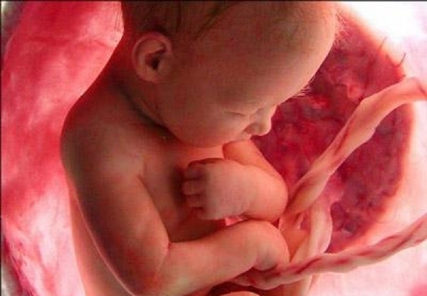
Amniotic fluid has the function of nourishing and protecting the fetus during pregnancy. The volume of amniotic fluid changes with each stage of fetal development. Through the survey of the volume of amniotic fluid, the doctor can assess the health status and some diseases of the fetus.
1. The role of amniotic fluid in pregnancy
Amniotic fluid is a nutrient-rich environment that nourishes the embryo, in addition, amniotic fluid also helps protect and protect the fetus from collisions and trauma, ensuring a sterile environment for the baby during pregnancy. amniotic sac. Amniotic fluid creates an environment for the fetus to develop harmoniously and to normalize its position in the mother's genital tract in the last months of pregnancy.
2. Amniotic fluid changes during pregnancy
3. Possible abnormalities in amniotic fluid volume
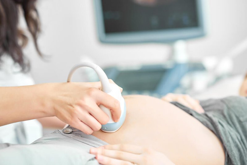
The amount of amniotic fluid increases gradually from 250 to 800ml in about 16 - 32 weeks, then stabilizes at this level until 36 weeks and then gradually decreases to about 500 ml when the due date comes. In fact, people use ultrasound to evaluate how little or too much amniotic fluid there is; and there are 2 ways of measuring: either directly measure any amniotic space (measurement of the maximum diameter of the cavity or measure the volume of the cavity) or divide the chamber from the arch into 4 parts and evaluate the amniotic cavity in all 4 parts , then added together to get the amniotic fluid index (AFI). Clinical assessment is based on abdominal palpation, which shows that the uterine height does not correspond to gestational age (smaller when there is less amniotic fluid or more when there is more amniotic fluid), whether the abdomen is firm or loose, but these signs are also present. It can be due to other reasons such as large or malnourished children, multiple pregnancies, too thin or too overweight. Therefore, ultrasound remains the most accurate assessment.
Possible abnormalities in amniotic fluid volume such as polyhydramnios, oligohydramnios, and amniotic fluid loss.
3.1 Polyhydramnios Polyhydramnios is an excess of amniotic fluid. Pregnant women are diagnosed with polyhydramnios when the amniotic fluid index (AFI) on ultrasound is more than 25 cm. Polyhydramnios is common in multiple pregnancies and some abnormalities of the fetal central nervous system such as hydrocephalus, anencephaly, meningocele, split spine....
About 2/3 of cases polyhydramnios is idiopathic. Causes of polyhydramnios include:
Maternal diabetes: Polyhydramnios is detected in 10% of pregnant women with diabetes, especially in the third trimester of pregnancy. When you have diabetes and your blood sugar is not well controlled, your baby may produce more urine. Therefore, lowering your blood sugar will reduce the amount of amniotic fluid. Mothers carrying twins or multiples: Polyhydramnios can occur because the metabolism between the two fetuses is not balanced (one fetus has less amniotic fluid while the other has more amniotic fluid). Causes in the fetus: The baby will stop the process of drinking amniotic fluid - urinating, leading to an excess of amniotic fluid. This condition occurs when malformations in the fetus such as cleft palate, pyloric stenosis... Other factors that may increase polyhydramnios are: Anemia in the fetus; fetal infection, blood type incompatibility between mother and baby... Effects of polyhydramnios on fetal health and development: polyhydramnios occurs in about 1% of pregnancy, polyhydramnios if due to malformations or deformed, the prognosis is very poor. Polyhydramnios makes the fetus easy to move in the uterus, so it can cause the umbilical cord to wrap around the neck, abnormally. Polyhydramnios causes the mother's abdomen to swell, making it difficult for pregnant women to breathe, causing uterine contractions and premature labor, which increases the baby's birth mortality rate. During labor, polyhydramnios can cause prolonged labor, causing fetal distress, and the mother has uterine atony, causing postpartum hemorrhage. Besides, polyhydramnios can cause sudden rupture of membranes and complications of sudden rupture of membranes are: placental abruption, umbilical cord prolapse, abnormal position and amniotic fluid embolism. These are life-threatening for both mother and child. Of course, these risks vary depending on the severity and cause of polyhydramnios.

In most cases of mild hyperhydramnios there is nothing to worry about. Your doctor will tell you to have regular checkups and give you some diuretics. If the cause of the infection can be determined, it can be treated with antibiotics that are safe for the fetus.
For severe polyhydramnios, there are signs of problems related to the fetus that need to be monitored. Your doctor will closely monitor your amniotic fluid volume, if it increases too quickly you may need surgery, amniocentesis to remove the amniotic fluid.
In addition, pregnant women need to regularly visit the doctor for monitoring, and may even have to be hospitalized and intervene as soon as necessary when symptoms such as shortness of breath and chest tightness appear; The abdomen enlarges rapidly and clearly, and the pain is sudden.
3.2 oligohydramnios, amniotic fluid oligohydramnios, a condition in which amniotic fluid is less than normal, when the amniotic index (AFI) is less than 5cm and the amniotic membrane is intact, common in abnormalities of the urinary system and fetal digestive system . Amniotic fluid is defined when the amniotic fluid index is less than 3cm. Low amniotic fluid is also common in maternal malnutrition, fetal malnutrition, pregnancy past due date, premature rupture of membranes, premature rupture of membranes....
Effects of oligohydramnios on fetal development: Depends on the situation Status, causes and gestational age to which the lack of amniotic fluid will affect more or less.
In case a pregnant woman has a lack of amniotic fluid in the first 3 months of pregnancy or a few weeks after, there is a possibility of miscarriage and stillbirth. At the same time, this will also affect the development and lung function of the fetus.
In case the mother has a lack of amniotic fluid in the last 3 months of pregnancy, most do not cause dangerous complications. The patient will be closely monitored the health of both mother and child and may be given water to replenish amniotic fluid for pregnant women. However, oligohydramnios during this stage can cause the fetal position to be reversed because there is not enough amniotic fluid needed to rotate the head downwards.
Lack of amniotic fluid can also cause the mother to have premature rupture of membranes. If the water breaks, causing oligohydramnios when not in labor or at the beginning of the birth, it will infect the amniotic fluid, thereby infecting the fetus, infecting the uterus... both mother and baby are dangerous to health and live. The doctor will consider the risk of amniotic fluid infection and the condition of the fetus to decide whether to intervene to help the mother deliver early.
Any abnormal change in amniotic fluid volume can adversely affect the development of the fetus, so during pregnancy, the mother needs to be periodically monitored to ensure the quality of amniotic fluid.
Doctor Nguyen Anh Tu has 6 years of experience in obstetrics and gynecology ultrasound, specially researched and trained in pregnancy ultrasound - prenatal diagnosis. Dr. Tu has completed courses on ultrasound - prenatal diagnosis of the FMF International Fetal Medicine Association; trained in consulting and implementing diagnostic intervention techniques in fetal medicine and participated in many specialized conferences and seminars on Fetal Medicine. Currently a doctor at the Department of Obstetrics and Gynecology, Vinmec Hai Phong International General Hospital
Để đặt lịch khám tại viện, Quý khách vui lòng bấm số HOTLINE hoặc đặt lịch trực tiếp TẠI ĐÂY. Tải và đặt lịch khám tự động trên ứng dụng MyVinmec để quản lý, theo dõi lịch và đặt hẹn mọi lúc mọi nơi ngay trên ứng dụng.
Dịch vụ từ Vinmec
-
 Sự hình thành và nguy hiểm của dây rốn thắt nút
Dây rốn là đường dẫn duy nhất kết nối mẹ và con, mang chất dinh dưỡng và dưỡng khí đến nuôi em bé. Tuy nhiên, khi dây rốn bị thắt nút lại sẽ gây ảnh hưởng nghiêm trọng đến thai ...
Đọc thêm -
 Chẩn đoán và xử trí dây rốn thắt nút
Theo các số liệu thống kê, tỷ lệ dây rốn thắt nút chiếm 0.3 – 2.2% các trường hợp mang thai và tỷ lệ tử vong của thai nhi gặp phải tình trạng này thường tăng cao gấp 4 lần ...
Đọc thêm -
 Thai nhi 38 tuần bị giảm cân có sao không?
Em đang mang thai 38 tuần. Lúc thai 37 tuần, em đi khám bé được 2900g, nay em khám định kỳ lại thai 38 tuần giảm còn 2750gr. Vậy bác sĩ cho em hỏi thai nhi 38 tuần bị giảm ...
Đọc thêm -
 Biến chứng nguy hiểm của xoắn dây rốn thai nhi
Dây rốn thai nhi là một trong những bộ phận quan trọng giúp thai nhi tiếp nhận oxy và nguồn dinh dưỡng từ mẹ. Một trong những vấn đề về dây rốn khá nguy hiểm đó là xoắn dây rốn. ...
Đọc thêm -
 Thai nhi 28 tuần bị giãn đài bể thận 2 bên có sao không?
Thai em được 28 tuần, siêu âm giãn đài bể thận 2 bên, bên trái 8,2mm, bên phải 8,4mm, không có triệu chứng khác bất thường. Vậy bác sĩ cho em hỏi thai nhi 28 tuần bị giãn đài bể ...
Đọc thêm