Brain Infarction: What you need to know
This is an automatically translated article.
1. What is cerebral infarction?
Cerebral infarction is a pathological process that reduces blood flow to an area of the brain due to narrowing, occlusion of a cerebral blood vessel or due to hypotension. Cerebral ischemia is a condition in which the blood supply to part of the brain is cut off. If the lack of blood supply is not corrected or persists, that part of the brain will die from lack of oxygen and glucose. This area of the brain that is damaged by a lack of blood supply is called a cerebral infarction.
Cerebral infarction accounts for about 80% of cerebral strokes, the remaining 20% is cerebral hemorrhage, subarachnoid hemorrhage. The annual incidence of cerebral infarction is relatively high, about 130/100,000 people/year.

2. Causes of cerebral infarction
Common causes of ischemic stroke are:
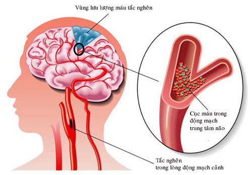
Atherosclerosis of large vessels accounts for 50%, of which large blood vessels outside the skull account for 45%, large blood vessels in the skull account for 5%. Causes from the heart cause blood clots such as open heart disease, atrial fibrillation, arrhythmias, heart failure ... creating blood clots go to the brain, accounting for 20%. Obstruction of small blood vessels in the brain, common in patients with hypertension, diabetes accounts for 25%. Non-atherosclerotic arterial disease accounts for < 5%. Blood diseases such as coagulopathy, blood cell disease, congenital abnormalities of blood vessels... accounted for <5%.
3. Diagnosis of cerebral infarction
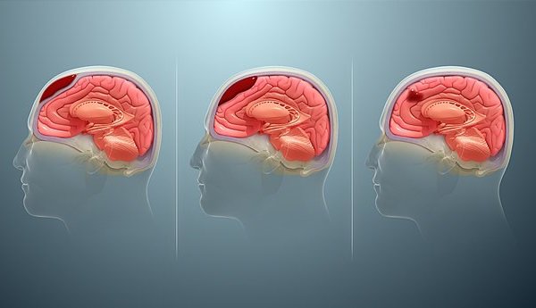
Symptoms of cerebral infarction occur suddenly while the patient is sleeping. Typical symptoms are headache, vomiting or nausea, hemiplegia. There may be disturbances of consciousness if the patient has extensive cerebral infarction, bilateral cerebral infarction, or brain stem infarction.
3.1 Subclinical diagnosis Brain CT scan: During the acute phase 3-6 hours after the onset of the disease, brain CT changes are very subtle, mainly due to cerebral edema in the ischemic area. caused by the brain.
Early signs of cerebral infarction on brain computed tomography include loss of gray matter white matter boundaries, sclerotic sulcus sulcus, stenosis of Sylvius cleft, loss of the insular band, ventricular stenosis and basal pool, and hyperattenuation. blood vessels in the area of the Willis polygon due to blood clots in particular the middle cerebral artery.

Brain magnetic resonance imaging: In the early stages of the disease, the cerebral infarction is slightly reduced on T1W images, increased on T2W images. Injecting contrast material into the body showed that the lesion was not enhanced. In the subacute phase (after 1 week of illness), the cerebral infarction is hypointense on T1-weighted images, and hyperintense on T2-weighted images. Intravenous contrast injection showed that the lesion was enhanced by the drug. In the late stage of the disease, the cerebral infarction is reduced in signal on T1-weighted images, and increased in signal on T2-weighted images. The mass effect usually disappears after 1 month, the contrast enhancement gradually subsides after a few months.
Digital brain angiography with background erasure: Digital brain angiography with background deletion (DSA) is an invasive technique aimed at accurately diagnosing cerebrovascular diseases and at the same time removing blood clots. through the intravascular route: after detecting the cerebral vascular occlusion through cerebral angiography by angiography and inserting an instrument that both pokes and sucks the clot out (aspirator, catheter).
4. Signs of a stroke
When you see a patient with FAST symptoms (English): distorted mouth, weak limbs, change in voice, need to go to the hospital immediately.
Some other stroke warning symptoms such as dizziness, headache, weakness in limbs, distorted mouth, slurred speech, coma, convulsions... When seeing these signs, the patient should be taken to the hospital soon to treat. Because if the treatment window is exceeded, the patient can be severely disabled if not treated in time.
5. Prevention of stroke recurrence

Treatment of causes of cerebral infarction: diseases of high blood pressure, diabetes, cardiovascular disease, dyslipidemia. Reasonable diet: Implement a diet to reduce fat, reduce salt (THA), reduce starch and sugar (DM), eat more green vegetables, exercise 30 minutes a day, stop smoking and alcohol and avoid obesity. Periodic re-examination and treatment according to the doctor's instructions When there are signs of suspicion of cerebral infarction, it is necessary to bring the patient to the hospital immediately. When there are symptoms suggestive of stroke such as weakness, distortion of the mouth, difficult to say, ..., the patient needs to quickly go to the hospital, absolutely do not handle it at home. Patients should be diagnosed early and accurately so that doctors will choose the most appropriate treatment. Time to hospital as soon as possible, especially within the first 6 hours after the stroke, will have the highest chance of recovery.
Currently, Magnetic Resonance Imaging - MRI/MRA is considered a "golden" tool for brain stroke screening. MRI is used to check the condition of most organs in the body, especially valuable in detailed imaging of the brain or spinal nerves. Due to the good contrast and resolution, MRI images allow to detect abnormalities hidden behind bone layers that are difficult to recognize with other imaging methods. MRI can give more accurate results than X-ray techniques (except for DSA angiography) in diagnosing brain diseases, cardiovascular diseases, strokes,... Moreover, the process MRI scans do not cause the side effects seen in X-rays or computed tomography (CT).
Vinmec International General Hospital currently owns a 3.0 Tesla MRI System, which is equipped with state-of-the-art equipment by GE Healthcare (USA) with high image quality, allowing for a comprehensive assessment, without omitting the injury without leaving any damage. and reduce shooting time. Silent technology helps to reduce noise, create comfort and reduce stress for the client during the shooting process, resulting in better image quality and shorter imaging time. With the state-of-the-art MRI system With the application of modern methods of cerebral vascular intervention, a team of experienced and well-trained neurologists and radiologists, Vinmec is a prestigious address for stroke risk screening and screening. reliable goods.
In the past time; Vinmec has successfully treated many cases of stroke in a timely manner, leaving no sequelae: saving the life of a patient suffering from 2 consecutive strokes; Responding to foreign female tourists to escape the "death door" of a stroke;...
Please dial HOTLINE for more information or register for an appointment HERE. Download MyVinmec app to make appointments faster and to manage your bookings easily.
-
 Nutrition for patients after stroke
Bệnh nhân sau đột quỵ bị ảnh hưởng nghiêm trọng về mặt sức khỏe. Bên cạnh các phác đồ điều trị và vật lý trị liệu, chế độ dinh dưỡng cho người sau tai biến vô cùng quan trọng. Thực ...
Readmore -
 Typical symptoms warning cerebrovascular malformations
Dị dạng mạch máu não gây ra nhiều vấn đề đối với sức khỏe mà phổ biến nhất là vỡ mạch dẫn đến đột quỵ do xuất huyết não hoặc gây áp suất tiếp giáp lên não dẫn đến động ...
Readmore -
 Intracranial hematoma is diagnosed by what medical technique?
Tụ máu nội sọ là khối máu tụ trong sọ nếu không được chẩn đoán phát hiện sớm có thể gây ra những biến chứng nguy hiểm. Những phương pháp chẩn đoán tụ máu nội soi như CT scan, chụp ...
Readmore -
 Magnetic Resonance Perfusion (MRI) - P2
MRI tưới máu não là kỹ thuật được sử dụng để đánh giá sự phân bố máu đến nhu mô não. Sử dụng kỹ thuật này giúp chẩn đoán phân biệt một số bệnh lý thần kinh và đánh giá ...
Readmore -
 Coarctation of the aorta
Coarctation of the aorta is a treatment method that is being widely applied thanks to its low-invasive advantages, high success rate, and very safe, especially for young children. As a result, patients avoid the risk of unpredictable complications such as ...
Readmore