Atelectasis: Diagnosis and treatment
This is an automatically translated article.
1. What is atelectasis?
Atelectasis can interfere with the airway, especially if there is additional lung disease. Treatment for this condition depends on the cause and severity of the atelectasis.
2. Diagnosis of atelectasis
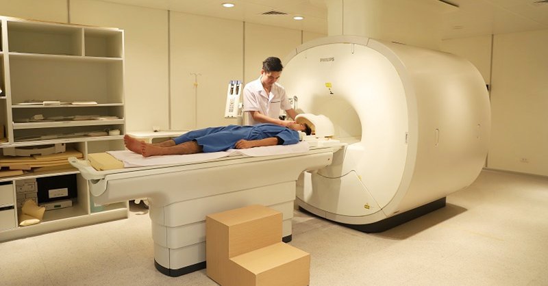
CT scan: Because CT is a more precise technique than X-ray, it can sometimes help better detect the cause and type of atelectasis. Oxygen measurement: This simple test uses a small device placed on your finger to measure the level of oxygen in your blood. It helps determine the severity of atelectasis. Thoracic ultrasound: This noninvasive test can help your doctor tell the difference between atelectasis, pulmonary sclerosis, pneumonia caused by fluid in the air sacs, and pleural effusion. Bronchoscopy: Using a flexible, lighted camera-tipped tube inserted down the throat allows the doctor to see what could be causing the blockage, such as a mucus plug, tumor, or foreign body. This method can also be used to remove blockages.
3. Treatment of atelectasis
3.1 Chest physiotherapy
Performing deep breathing exercises (spirometry) and using a device to assist with a deep cough can help clear secretions and increase lung volume. Position the body so that the head is lower than the chest (postural drainage). This allows the mucus to better drain from the bottom of the lungs. Tap the chest over the collapsed area to loosen the mucus. This technique is called the typing method. You can also use mechanical mucus cleaning devices, such as an air pulse vibrating vest or hand tools.
3.2 Surgery
If a tumor is causing atelectasis, treatment may include surgery to remove or shrink the tumor, with or without other cancer treatments (chemotherapy or radiation).

3.3 Breathing treatment
For children, childhood trauma is often caused by airway obstruction. To reduce the risk of illness, keep small objects out of the reach of children.
In adults, anemia often occurs after major surgery. If surgery is planned, talk to your specialist about strategies to reduce risk. Some research suggests that breathing exercises and muscle training may reduce the risk of disease after certain surgeries. In addition, smoking is also recommended because smoking increases mucus production and affects hair-like structures in the bronchial tubes (hairs).
Please dial HOTLINE for more information or register for an appointment HERE. Download MyVinmec app to make appointments faster and to manage your bookings easily.
-
 Uses of Zencombi
Zencombi is used to relax bronchial muscles and increase air volume in patients with airway obstruction. Zencombi combines two drugs according to different mechanisms, so it has a synergistic effect in relaxing bronchial muscles.
Readmore -
 Uses of Actidose
Actidose or activated charcoal is a drug in the detoxification group. However, the use and safety of the drug needs to be carefully examined. Hopefully the following information will help you understand and limit the dangers of taking Actidose.
Readmore -
 Uses of Tolbupas
Tolbupas is indicated for the relief of symptoms of shortness of breath caused by obstructive airway disease in patients with acute bronchitis, bronchial asthma, pneumothorax, chronic bronchitis... Let's learn about the uses. , notes when using Tolbupas through the article ...
Readmore -
 Swallowing batteries in children - What you should know
Bé nuốt pin nếu không được xử lý kịp thời sẽ đe doạ đến tính mạng. Thậm chí nhiều phương pháp truyền miệng lấy pin bị mắc kẹt trong cơ thể của trẻ không những không có tác dụng mà ...
Readmore -
 Endoscopic tracheobronchial stent placement
Đặt stent khí – phế quản qua nội soi là gì, những trường hợp nào cần thực hiện kỹ thuật này, trường hợp nào chống chỉ định, quy trình đặt stent diễn ra như thế nào, những tai biến nào ...
Readmore