Echocardiography to evaluate and diagnose tumors in the heart
This is an automatically translated article.
1. Overview of tumors in the heart
Tumors in the heart can be benign or malignant. Primary malignancies of the heart are rare, mostly benign, with an incidence of 75% of all cases. In addition, there are cases of melanoma metastasized from other organs to the heart.
1.2 Common Heart Tumors Since heart tumors are mostly benign, this section will detail the common types of benign intracardiac tumors. They are:
Mucinous tumor: The most common benign tumor, located in the heart cavities. Most mucinous tumors occur in the left atrium; Fibroids: Grow in the myocardium or in the endocardium. Fibrous tumors tend to occur on the heart valves, which may be related to an inflammatory response; Rhabdomyomas: usually develop in the myocardium or in the endocardium. Rhabdomyomas are common in young children and are associated with tuberous fibrosis, renal tumors, sebaceous gland tumors of the skin, and cardiac arrhythmias. Rhabdomyolysis tends to occur in the ventricular septum; Teratoma: Tumor develops from embryonic germ cells. The teratoma of the pericardium often adheres to the base of the great vessels, is common in children, and is less common in the form of cysts or lipomas. Teratoma is usually asymptomatic, mainly detected through routine chest and chest x-ray; Lipomas: Can originate in any tissue of the heart, most commonly in children. 1.3 Symptoms of Tumors in the Heart When heart tumors are small, they usually don't cause symptoms. As tumors grow, they will occupy a large volume in the heart chambers, can descend into the ventricles, making the patient short of breath, rapid heart rate and the patient will be more comfortable when lying on the right side. Sometimes, a heart tumor can block the mitral orifice, preventing blood from reaching the left ventricular chamber, causing fainting or sudden death.
Tumors also often move according to the heart's contraction cycle, so they are easy to break into small pieces. Tumor fragments drifting through the blood stream can cause stroke, embolism of the limb. These are very serious complications of heart tumours.
Metastatic melanoma from other places to the heart is also very dangerous because it can cause congestive heart failure, arrhythmia and heart block, pericardial effusion, ... with a very poor prognosis and risk of death. high mortality.

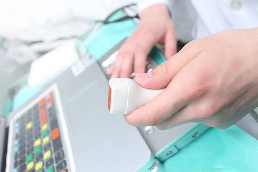
2. Ultrasound - an effective method of diagnosing tumors in the heart
Echocardiography is usually done by 3 methods:
Stress echocardiography: Performed when the doctor conducts an electrocardiogram or gives the patient medication that can make the heart beat faster; Doppler ultrasound: Measures blood flow velocity at locations in the heart chambers; Transesophageal ultrasound: The patient has to swallow a probe with a thin optical cable connected to the ultrasound machine for the doctor to observe, diagnose and detect the disease. On echocardiography, the doctor can clearly see the tumor moving in the heart chambers, the tumor stalk is attached to the heart wall. In addition, this imaging technique also allows doctors to accurately assess the nature and size of heart tumors, see the tumor's movement through the mitral valve during the heart's contraction cycle. Therefore, it is considered to be the best diagnostic test in the evaluation of patients with intracardiac tumors.
Echocardiography is an important imaging method to help detect lesions and tumors in the heart. However, in some cases, it is necessary to combine with other imaging methods such as CT scan, MRI scan,... to better evaluate the characteristics of the tumor.
To protect heart health in general and detect early signs of myocardial infarction and stroke, customers can sign up for Cardiovascular Screening Package - Basic Cardiovascular Examination of Vinmec International General Hospital . The examination package helps to detect cardiovascular problems at the earliest through tests and modern imaging methods. The package is for all ages, genders and is especially essential for people with risk factors for cardiovascular disease.
Please dial HOTLINE for more information or register for an appointment HERE. Download MyVinmec app to make appointments faster and to manage your bookings easily.
-
 Is there a need to treat 1/4 heart valve regurgitation and dangerous disease?
Hi doctor! My wife went for an echocardiogram at the hospital and found that there was a 1/4 heart valve regurgitation. The doctor asked me, do I need to treat 1/4 heart valve regurgitation and dangerous disease? I sincerely thank.
Readmore -
 Is there a chance for a heart tumor to be cured or surgically removed?
I have a newly discovered heart tumor. So let me ask, is there a chance for a heart tumor to be cured or operated on, doctor?
Readmore -
 Advantages and limitations of ultrasound machines in diagnostic imaging
Siêu âm là phương pháp chẩn đoán hình ảnh hiện đại được thực hiện trong các khâu thăm khám, chẩn đoán, phục vụ công tác điều trị bệnh tại các cơ sở y tế, bệnh viện. Vì thế, hiện nay ...
Readmore -
 The role of fetal Doppler ultrasound
Kỹ thuật siêu âm Doppler cơ tử cung là một trong những phương pháp siêu âm thai tiền sản có vai trò quan trọng và rất cần thiết trong sản khoa. Siêu âm Doppler thai giúp theo dõi và đánh ...
Readmore -
 Cerebral artery ischemic stroke in children: Clinical and diagnostic
Đột quỵ thiếu máu cục bộ động mạch não ở trẻ em hiện là một bệnh lý vô cùng nguy hiểm và ảnh hưởng nghiêm trọng tới sức khỏe nếu không được phát hiện và can thiệp kịp thời. Do ...
Readmore