Magnetic resonance imaging of the orbit and optic nerve
This is an automatically translated article.
1. What is the purpose/meaning of the technique?
Injury to the orbit includes many different pathologies, from diseases of the eyeball, pathologies around the eyeball to diseases in the brain affecting the path of the optic nerve; This has created challenges in diagnosis, management, and treatment.
With the development of the imaging industry, especially the introduction of magnetic resonance imaging (MRI), has helped to evaluate soft tissue in detail, allowing better characterization of lesions.
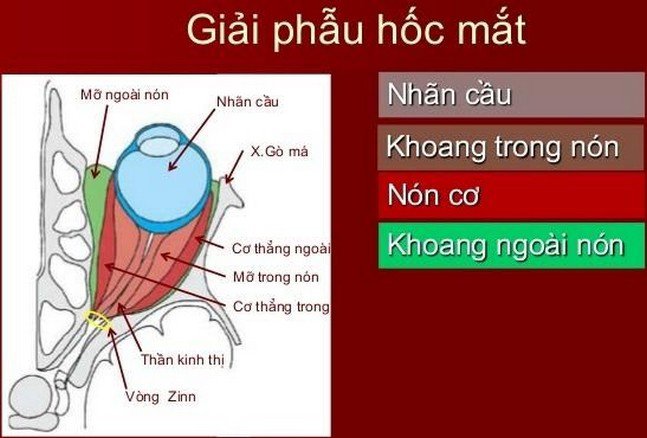
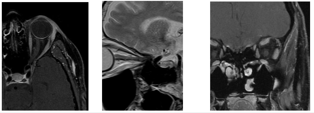
Magnetic resonance imaging of the orbit and optic nerve with injection of magnetic contrast agent is a modern technique, widely applied in the diagnosis of eye diseases, diseases of the ophthalmic muscles, lesions in the eyes. cones (such as vascular abnormalities, inflammation or optic neuroma...); as well as extra-conical lesions (such as abscesses, cord tumors, and bone lesions...). It has high resolution to distinguish pathological structures from normal structures such as eyeball, optic nerve, oculomotor muscles...
Therefore, orbital magnetic resonance imaging plays an increasingly important role. , especially in the event of an inadequate history and clinical assessment.
2. Indications/contraindications?
2.1 Indications Manifestations of visual loss on clinical examination are consistent with intracranial lesions before or after optic interference Abnormalities of blood vessels of the orbit (such as arteriovenous malformation, carotid-cavernous fistula, orbital varicose veins). History of visual disturbances Meningiomas of the optic nerve Thyroid eye disease Optic neuritis Tumor or suspected optic nerve glioma Tumor intraorbital abscess Protrusion of the eyeball on one eye
2.2 Contraindications Patients with pacemakers (absolute contraindications) In people with magnetic metals (relative contraindications), especially with metallic foreign bodies inside the eye sockets People who are afraid of lying alone Inability to lie still Women under 12 weeks pregnant
3. How to do it
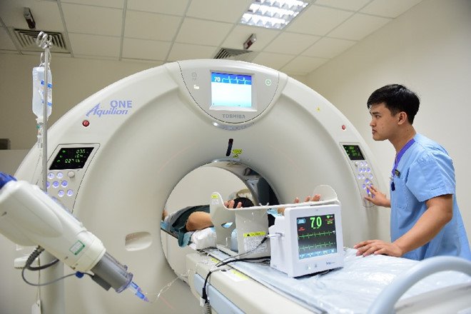
Patients who have an appointment should arrive at the MRI room 15 minutes before. When entering the Radiology Department, the patient will be thoroughly explained about the procedure to coordinate well with medical staff, check for contraindications, then change into the clothes of the MRI room and remove the objects. contraindicated use. The patient lies supine on the table, with the head placed on the coil and held still, the patient may be provided with an eye mask to help prevent eye movement during the scan.

When taking the patient's orbital magnetic resonance imaging, two-stroke imaging will be taken. The first stage is conventional cranial magnetic resonance imaging and localized orbital imaging, the second is localized imaging of the orbital area injected with magnetic contrast. The commonly used contrast agent is Dotarem, which makes it easy to tell the difference between normal tissue and a tumor, infection, or other abnormality. The normal brain magnetic resonance imaging time is about 20-25 minutes and the additional time for additional pulses for the eye socket and contrast injection is 30 minutes. The results of the orbital magnetic resonance imaging will be sent to the doctor for indication, then the doctor will explain the results to the patient.
4. Advantages/disadvantages
Advantages: Magnetic resonance has advantages over other imaging techniques, it has high image resolution, helps to detect very small lesions, even tumors <3mm in diameter. Cons: Like the general limitations of magnetic resonance imaging, orbital magnetic resonance imaging is not available in patients with pacemakers. Restricted in patients with metal in the body, patients in the first trimester of pregnancy, and claustrophobia.
5. Special points/advantages when performing techniques at Vinmec hospital
Vinmec Nha Trang International General Hospital has invested in a US-made 3.0 Tesla magnetic resonance imaging machine with an imaging room that meets the requirements of the Ministry of Health. This is a new step in healthcare, helping patients to access the most modern technology, save costs and diagnose diseases quickly and accurately. Patients with orbital magnetic resonance imaging at Vinmec Nha Trang International General Hospital not only have their results read carefully by their doctors, but also receive dedicated advice on risk factors and pathologies to have a better solution. the best care for your own health.
Vinmec International General Hospital is one of the hospitals that not only ensures professional quality with a team of leading medical doctors, modern equipment and technology, but also stands out for its examination and consultation services. comprehensive and professional medical consultation and treatment; civilized, polite, safe and sterile medical examination and treatment space.
Customers can go directly to Vinmec Nha Trang to visit or contact hotline 0258 3900 168 for support.
-
 Understanding neurofibromatosis type 2 (NF2)
U sợi thần kinh loại 2 là một trong những bệnh di truyền u sợi thần kinh do rối loạn nhiễm sắc thể 22 gây ra. Mặc dù lành tính nhưng khối u có thể ảnh hưởng đến khả năng ...
Readmore -
 Computed tomography of the brain without contrast for the diagnosis of intra-axial brain tumors
Chụp cắt lớp vi tính sọ não không tiêm thuốc đối quang là kỹ thuật hình ảnh được áp dụng rộng rãi trong chẩn đoán các bệnh lý thần kinh. Chụp cắt lớp vi tính sọ não được chỉ định ...
Readmore -
 Learn about tumors in the skull
Các khối u trong sọ có thể liên quan đến não hoặc các cấu trúc khác như dây thần kinh sọ, màng não. Các khối u thường phát triển trong giai đoạn đầu hoặc giữa tuổi trưởng thành nhưng có ...
Readmore -
 Common symptoms warning brain tumor
Some of the symptoms of a brain tumor are the same as those of other medical conditions. Diagnosis of brain tumor requires a combination of physical examination and laboratory tests. The following article will mention some of the common symptoms ...
Readmore -
 Treatment of brain tumors that recur after surgery
Tình trạng u não tái phát sau mổ không phải là hiếm gặp mà còn rất phổ biến, ngay cả khi phẫu thuật thành công. Do đó, việc chuẩn bị sẵn sàng cho tình trạng này cũng như xem xét ...
Readmore