What is “atypical hyperplasia” in the pathology of a breast biopsy?
This is an automatically translated article.
The article is written by MSc, Doctor Nguyen Van Khanh, Laboratory Department - Vinmec Times City International Hospital
After you have had a breast biopsy, the specimen will be studied under a microscope by a pathologist. The pathologist will then send a report of the results to your treating physician. The information in this report will be used to help manage your care. The questions and answers below will help you understand medical terms you might see in a breast biopsy pathology report, such as a fine needle biopsy or an open biopsy.
In fine-needle biopsies, a needle is used to collect a specimen in the abnormal area. An open biopsy removes the entire abnormal area and is usually accompanied by a small amount of surrounding normal tissue. An open biopsy is like a type of breast-conserving surgery called a lumpectomy.
1. What does overproduction mean?
Normal mammary gland tissue is made up of ducts with a group of sacs called lobules at one end. Hyperplasia is the term used when there is growth of cells inside the ducts and/or lobules of the mammary gland. Hyperplasia is not cancer. Normally, the ducts and the lobules are lined by two layers of cells. Hyperplasia means there are more than two layers of cells. If the developed component looks similar to the normal component under the microscope, the hyperplasia can be termed as normal hyperplasia. Some cases have a more unusual growth-like component, which can be called atypical hyperplasia.
There are two main types of mammary gland hyperplasia: ductal hyperplasia and lobular hyperplasia. What constitutes tubular hyperplasia or lobular hyperplasia is based on the microscopic appearance of the cells rather than on the appearance of ducts or lobules.

2. Does your report mention E-cadherin, what does that mean?
E-cadherin is a test that the pathologist can use to determine whether the hyperplasia is tubular or lobular (cells in atypical lobular hyperplasia are often negative for E-cadherin) . If your report does not mention E-cadherin, it means that this test is not needed to diagnose you with this type of hyperplasia.
3. What is the meaning of atypical tubular hyperplasia?
In atypical hyperplasia, cells grow abnormally and have some (but not all) features of ductal carcinoma in situ (which is a precancerous lesion). This means that atypical ductal hyperplasia is not a precancerous lesion, although it is associated with an increased risk of breast cancer later in life.
If atypical ductal hyperplasia is found on a needle biopsy, it is necessary to obtain multiple tissue samples in that area to ensure that no more serious lesions are present in the breast. The removed tissue sample is viewed under a microscope, and if there is no more serious damage, no further treatment is necessary. Patients only need to be followed up by clinical breast examination and by diagnostic breast imaging tests such as mammography.
If atypical hyperplasia is found on an open biopsy, no further surgery is necessary, but your doctor may recommend additional medications to reduce your breast cancer risk.

4. What is the meaning of atypical lobular hyperplasia?
Atypical lobular hyperplasia, also an abnormal growth of cells in the lobules of the breast, is associated with an increased risk of breast cancer. If atypical lobular hyperplasia is detected on a needle biopsy, some physicians are of the opinion that further surgery is needed to make sure there are no other serious injuries, while others are Doctors suggest that only follow-up with a physical breast exam and imaging tests (eg, mammograms) is needed. If atypical lobular hyperplasia is found on an open biopsy (mammary-conserving surgery), the patient is most likely to be observed without further treatment, but your doctor may recommend oral medications. Add drugs to reduce the risk of breast cancer.
5. What do special tests such as: high molecular weight cytokeratin, CK903, CK5/6, p63, MSA, light chain SMM, calponin or keratin mean?
These special tests are sometimes used by pathologists to help make the correct diagnosis for a variety of breast lesions. They are both immunohistochemical tests performed on your biopsy specimen. Results are available in 2-3 days. Whether or not these tests are mentioned in your results has nothing to do with the accuracy of the diagnosis.

6. What does this mean if your results use any of the following terms: common ductal hyperplasia, glandular disease, fibroadenoma, scleral scar, complex fibrous lesion, papilloma? , apocrine metaplasia, cyst, columnar cell change, collagen bridge, ductal dilatation, cystic fibrosis change.
All of these terms refer to benign (noncancerous) changes in the breast that the pathologist can see under the microscope. They are not important when seen on a biopsy specimen containing atypical tubular hyperplasia or atypical lobular hyperplasia.
7. What does this mean if your results refer to microcalcifications or calcifications.
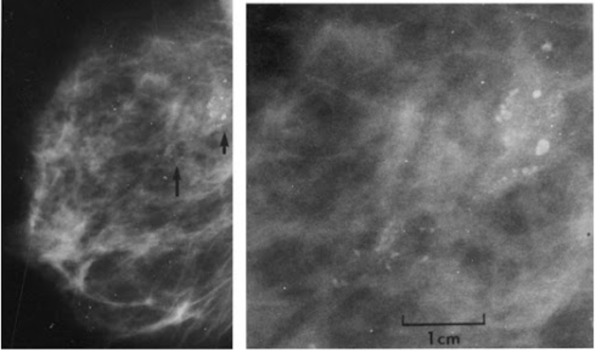
Microcalcifications or calcifications are calcium deposits found in noncancerous and cancerous lesions of the mammary gland. They can be detected both on mammograms and under the microscope. Because calcifications can be found in areas containing cancer, the presence of calcifications on mammograms may be an indication that a biopsy of the calcified area is needed. Then, when the biopsy is done, the pathologist looks at the removed tissue sample to make sure it contains calcifications. If there is calcification, the treating physician understands that the tissue sample was taken in the correct area (the abnormal area containing calcification was detected on the mammogram).
At Vinmec International General Hospital, there is a Breast Cancer Screening Package, which helps customers screen and detect breast cancer early even when there are no symptoms. When registering for the Breast Cancer Screening Package, customers will receive:
Examination and consultation with an oncologist. Breast cancer screening by bilateral breast ultrasound and mammogram. Up to now, Vinmec has become a prestigious address in breast cancer screening with:
Team of highly qualified and experienced doctors. Comprehensive professional cooperation with domestic and international hospitals: Singapore, Japan, USA, etc. Comprehensive treatment and care for patients, multi-specialty coordination towards individualizing each patient. Having a full range of specialized facilities to diagnose the disease and stage it before treatment: Endoscopy, CT scan, PET-CT scan, MRI, histopathological diagnosis, gene-cell testing, .. There are full range of mainstream cancer treatment methods: surgery, radiation therapy, chemotherapy, stem cell transplant..
Please dial HOTLINE for more information or register for an appointment HERE. Download MyVinmec app to make appointments faster and to manage your bookings easily.
References: American Cancer Society
-
 Can ultrasound detect thyroid cancer?
Siêu âm không thể phân biệt hoàn toàn ung thư tuyến giáp với các bệnh lý tuyến giáp khác. Điều này là do siêu âm không thể quan sát ở mức độ tế bào như sinh thiết và cho hình ...
Readmore -
 How to preserve samples and conduct pathology
Xét nghiệm giải phẫu bệnh học là công tác tiếp nhận, lấy mẫu bệnh phẩm, nhuộm tiêu bản, đọc các tiêu bản giải phẫu bệnh, bảo quản mẫu, hội chẩn bởi bác sĩ giải phẫu bệnh nhằm đưa ra chẩn ...
Readmore -
 Stages of thyroid cancer
Cancer stage describes the growth in size and spread beyond the organ in the body far away from where the cancer cells started. Different types of thyroid cancer go through different and often complex stages. This article provides detailed information ...
Readmore -
 What are the pathologic results of a prostate cancer biopsy?
Prostate cancer is cancer that develops in the prostate gland in men. These cancer cells become more dangerous when they spread to other organs, especially to the bones or to the lymph nodes. Therefore, it is important to screen for ...
Readmore -
 How will cytology and biopsy specimens be handled?
Các quy trình và các phương pháp chuẩn được sử dụng để xử lý gần như tất cả các loại mẫu sinh thiết. Các quy trình này là các cách thông thường mà mẫu bệnh phẩm được chuẩn bị trong ...
Readmore