Ultrasound combined with intrauterine infusion Sonohysterography
This is an automatically translated article.
Article written by Dr. Nguyen Quang Nam, Department of Obstetrics and Gynecology, Vinmec Times City International General Hospital
1. What is ultrasound combined with intrauterine infusion?
Sonohysterography is a special type of ultrasound test. Fluid is introduced into the uterus through the cervix using a thin plastic tube. Sound waves are then used to create an image of the lining of the uterus. Liquids help show more detail than when ultrasound is used alone. This test can be done at a hospital or an obstetrician-gynecological clinic. It usually takes less than 30 minutes.
2. When is it necessary to combine ultrasound with uterine pump?
After the gynecological ultrasound doctor suspects endometrial diseases but it is not clear, the doctor will do ultrasound with water pump to clarify the lesions.

Abnormal growth inside the uterus such as: uterine polyps, mucosal hyperplasia, uterine adhesions; Scar tissue inside the uterus; Abnormal uterine shape; Tumor in the uterus.
3. What is done to prepare for the ultrasound?
A hysterosalpingogram is not done if you are or could be pregnant or if you have a pelvic infection. You may have a blood or urine test to rule out pregnancy before the procedure.
The test is usually done after the end of the period but before ovulation.
You will have a pelvic exam before the ultrasound, if your doctor thinks you have an infection you may need antibiotics to clear up the infection before the procedure.
If you have abnormal bleeding that is persistent or the bleeding does not go away, you may be given medicine to stop the bleeding before the test.

4. The main steps of ultrasound combined with intrauterine infusion
Sonohysterography has three main steps:
Perform a transvaginal ultrasound exam Insert fluid into the uterus Repeat an ultrasound exam.
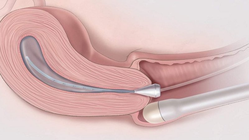
5. How is fluid inserted inside the uterus for an ultrasound?
After the first transvaginal ultrasound examination, the transducer is removed. A speculum is placed in the vagina. It keeps the vagina open. A swab is passed through the speculum to clean the cervix.
Next, a thin tube is inserted into the vagina and placed at the opening of the cervix or inside the uterus. Then the speculum is removed.
The probe is again inserted into the vagina. A sterile liquid is pumped slowly through the tube.
When the uterus is filled with fluid, the uterine cavity will dilate, the two mucosal edges separate, which is a condition for the doctor to clearly observe the lesion.
If the doctor replaces it with a special liquid containing safe microfoams, the fallopian tubes can be visualized on both sides. (Hycosy Ultrasound)
6. What are the common phenomena after ultrasound?

Most women are able to go home immediately and return to their normal activities that day. Some of the following symptoms may occur after the test:
Cramps Spots of blood or light bleeding Watery
7. What are the risks of ultrasound?
This test is very safe, but there is a rare risk of pelvic infection. Call your Doctor if you have any of the following symptoms:
Pain or fever for a day or two after you go home Abnormal vaginal discharge The results of an ultrasound are highly dependent on your expertise. technicians and equipment. Therefore, you should go to a reputable medical facility to do it. Vinmec International General Hospital is highly appreciated for the quality of imaging techniques, including ultrasound combined with intrauterine infusion.
A team of well-trained, professional, highly qualified and experienced technicians and doctors, regularly updated knowledge about new and advanced imaging techniques in the world world, helping to make the most accurate diagnosis for patients.
Modern equipment, completely imported from the UK, France, USA, Singapore, Japan, Korea, brings clear images and accurate results.
Please dial HOTLINE for more information or register for an appointment HERE. Download MyVinmec app to make appointments faster and to manage your bookings easily.
-
 Hysteroscopy polypectomy
Bệnh Polyp buồng tử cung nếu không được phát hiện và điều trị kịp thời có thể dẫn đến những biến chứng nguy hiểm như vô sinh, hiếm muộn, tăng nguy cơ bị buồng trứng đa nang, ung thư cổ ...
Readmore -
 Endometrial polypectomy procedure
Polyp buồng tử cung của mỗi bệnh nhân có kích thước khác nhau, có thể từ vài mm đến vài cm. Hiện nay, mổ nội soi là phương pháp phổ biến để điều trị polyp buồng tử cung. Phương pháp ...
Readmore -
 Issues to know about uterine pump ultrasound
Phương pháp này an toàn, không sử dụng tia xạ. Ngoài quan sát buồng tử cung có thể đánh giá gián tiếp ống dẫn trứng, các bất thường của tử cung, buồng trứng.
Readmore -
 Signs and diagnosis of uterine polyps
Đa số polyp buồng tử cung đều là lành tính, tuy nhiên, có thể ảnh hưởng đến khả năng sinh sản, sinh hoạt hằng ngày và làm giảm chất lượng cuộc sống của người bệnh. Bài viết này sẽ cung ...
Readmore -
What you need to know about hysteroscopy using physiological saline and bipolar cauterization
Phương pháp này thường được sử dụng trong thăm khám phụ khoa để giúp chẩn đoán và tìm ra nguyên nhân các bệnh lý về tử cung. Nội soi buồng tử cung từ rất lâu cũng đã khẳng định được ...
Readmore