Cells are the basic living units of the body. They form tissues, tissues form organs, and organs constitute the body. When cells are damaged, the body exhibits pathological symptoms. Cellular degeneration can either be reversible or irreversible, leading to various consequences.
1. Cellular Functions
Cells interact with their environment through two processes: anabolism and catabolism.
• Anabolism involves cells absorbing substances from the environment and converting them into compounds beneficial for cell survival and development.
• Catabolism refers to the breakdown of absorbed substances into metabolites (growth factors, proteins, etc.) and releasing these products into the environment.
Under normal circumstances, anabolism and catabolism maintain a state of balance. When this balance is disrupted, cells become damaged, and the body manifests diseases.
2. Causes of Cellular Damage
• Physical agents: Trauma, burns, radiation.
• Chemical agents: Drugs, chemicals.
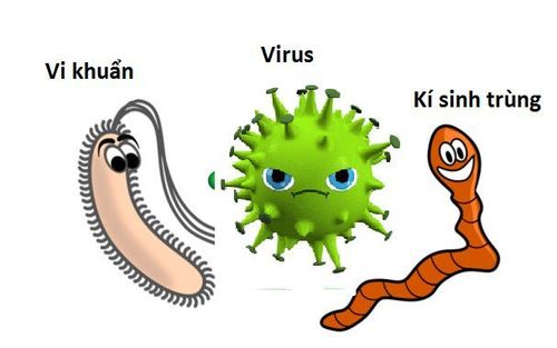
• Microorganisms: Bacteria, viruses, parasites.
• Immune responses.
• Genetic disorders.
• Nutritional imbalances.
3. Types of Cellular Damage
There are three types of cellular damage:
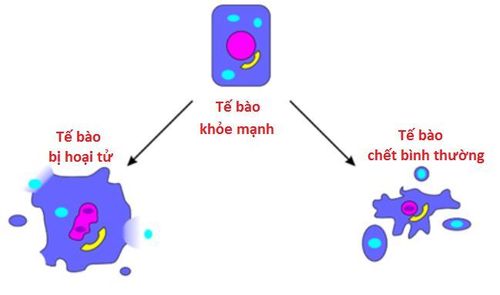
• Reversible damage (Cellular degeneration): Damage that may recover if pathological stimuli are reduced or removed.
• Irreversible damage (Cellular necrosis): Permanent damage leading to cell death.
• Adaptive damage: Including atrophy, hypertrophy, hyperplasia, hypoplasia, metaplasia, and dysplasia.
4. What is Cellular Degeneration?
Cellular degeneration refers to pathological changes in cell structure and function, primarily affecting the cytoplasm. This damage may reverse under favorable conditions. There are three main types of degeneration:
Granular degeneration:
The cell swells with water, and small red-stained granules appear in the cytoplasm when stained with hematoxylin-eosin (H.E.).
Functionality is reduced, and this nonspecific damage often occurs in parenchymal cells (e.g., liver cells during heart failure, renal tubular cells during toxicity).
Hydropic degeneration:
Closely related to granular degeneration, this involves swelling with water retained in intracellular vesicles, forming uneven clear spaces.
Common in parenchymal cells (e.g., liver or renal tubular cells) due to hypoxia or toxicity.
Fatty degeneration:
Fat droplets accumulate in the cytoplasm, appearing as large, round, clear vacuoles in H.E. staining.
Typically observed in liver cells, especially in the central lobular area, due to metabolic disorders (e.g., alcoholism, post-hepatitis).
5. What is Cellular Necrosis?
Cellular necrosis is the death of cells and tissues in a living organism. There are five main types of necrosis:
Coagulative necrosis:Necrotic tissue solidifies with intracellular and extracellular fluid coagulation, becoming friable and yellow-gray.
Common in limbs, fingers (vascular occlusion), and solid organs like the heart and liver (e.g., myocardial infarction).
• Liquefactive necrosis:
Necrotic tissue softens and liquefies, often with bacterial infiltration and inflammatory cells.
Seen in cerebral infarction (brain softening) and infected myocardial infarction.
• Caseous necrosis:
Necrotic tissue appears yellow-white, crumbly, and resembles cheese.
• Frequently associated with pulmonary tuberculosis and fungal infections (e.g., histoplasmosis).
• Fat necrosis:
Necrotic areas appear white, resembling candle wax, due to enzymatic fat digestion producing glycerol and free fatty acids.
Observed in acute pancreatitis, where pancreatic enzymes digest surrounding tissue.
• Fibrinoid necrosis:
Necrotic areas stain pink and resemble fibrin under eosin staining.
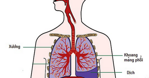
Often found on serous membranes (e.g., pleura, pericardium, peritoneum), causing roughened and adhesive surfaces.
6. What is Adaptive Damage?
There are four types of adaptive cellular damage:
• Changes in cell size:
Atrophy: Reduced cell size.
Hypertrophy: Increased cell size.
• Changes in cell number:
Hyperplasia: Increased cell number.
Hypoplasia: Reduced cell number.
• Changes in cellular differentiation:
Metaplasia: Replacement of one cell type with another.
Dysplasia: Abnormal cell growth or differentiation.
• Changes in cell metabolism:
Accumulation or retention of substances.
To arrange an appointment, please call HOTLINE or make your reservation directly HERE. You may also download the MyVinmec app to schedule appointments faster and manage your reservations more conveniently.
To arrange an appointment, please call HOTLINE or make your reservation directly HERE. You may also download the MyVinmec app to schedule appointments faster and manage your reservations more conveniently.