Acute pulmonary edema is common in patients with acute or chronic heart disease. I f acute pulmonary edema is detected early, it can save the patient's life. The doctor will have appropriate diagnosis and treatment methods.
1. Acute pulmonary edema
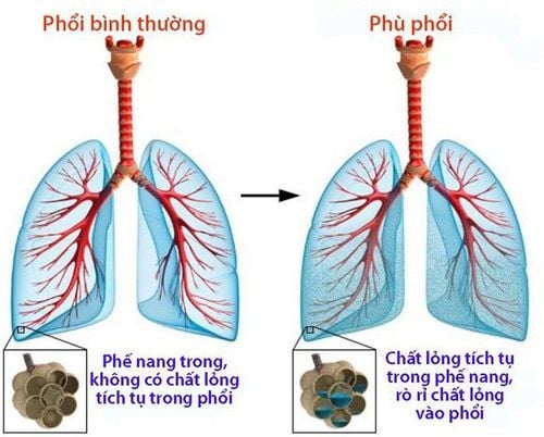
Acute pulmonary edema is a condition of acute suffocation caused by many different reasons, which cause too much water to escape from the pulmonary capillaries, causing pulmonary edema. Acute pulmonary edema is an acute disease that threatens life and can only be saved if early and effective intervention is given.
Acute pulmonary edema progresses in 3 stages: capillary phase, interstitial phase and alveolar phase. Clinically, acute pulmonary edema corresponds to the alveolar phase. Clinical manifestations are left heart failure and respiratory failure.
Left ventricular failure causes increased left atrial pressure, increased pulmonary venous and capillary pressure, increasing capillary permeability, resulting in fluid seepage into the alveoli, hindering gas exchange and respiratory failure.
2. Causes
Due to the imbalance of water exchange between pulmonary capillary tissues, alveoli and interstitial tissues including:
- Main factors: increased pulmonary capillary pressure, and increased capillary permeability.
- Favorable factors: decreased plasma colloid pressure, and obstruction of the lymphatic system.
Depending on the mechanism and cause of the pathogenesis, it is divided into 2 types: acute hemodynamic pulmonary edema and acute damage pulmonary edema.
- Acute hemodynamic pulmonary edema: is a condition of sudden increase in fluid pressure in the capillaries causing plasma to escape into the interstitium and alveoli without anatomical damage to the alveoli. Hemodynamic pulmonary edema is often caused by cardiovascular diseases including heart valve disease, hypertensive crisis, myocardial infarction, and myocarditis. Extracardiac causes such as acute and chronic glomerulonephritis, when performing procedures to drain pleural fluid too quickly or infusing too much or too quickly.
- Acute damage pulmonary edema: the leakage of plasma through the alveolar-capillary membrane without increased fluid pressure in the capillaries. Acute damage pulmonary edema is caused by bacterial, viral, and parasitic infections: bronchopneumonia, pneumococcal lobar pneumonia, pneumonic plague, malignant influenza, and malignant malaria. Acute poisoning: caused by inhalation of toxic substances such as CO2, NO2, SO2, pesticides, strong acids such as gastric juice, corrosive substances, and kerosene, drowning, hypoproteinemia, allergies, anaphylactic shock during blood transfusion.
3. Diagnosis
3.1 Medical history
- Medical history: rheumatic heart disease, congenital heart disease, chronic kidney disease
- Medical history of oliguria, hematuria and edema, the doctor may suggest glomerulonephritis
- Sudden heart failure should be considered myocarditis
- Rapid infusion
3.2 Tests
The patient needs to do some tests including:
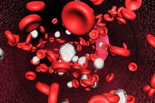
- Blood count
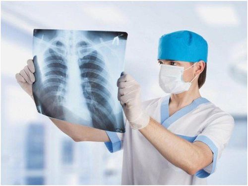
- Chest X-ray
- Blood gases
- Electrocardiogram (ECG)
- Echocardiography to diagnose heart disease, and assess myocardial contractility
- Urine analysis if acute glomerulonephritis is suspected
- If rheumatic heart disease is suspected: VS, ASO
- Troponin, high central venous pressure in case of suspected myocarditis
3.3 Differential diagnosis
- Pneumonia: cough, fever, dyspnea, crackles, chest X-ray showing lung consolidation.
- Asthma attack: history of asthma attack, wheezing in the lungs, chest X-ray showing pulmonary edema.
- Pulmonary hemorrhage: bloody sputum, no signs of heart failure, no pulmonary edema on X-ray.
- Acute respiratory distress syndrome: pulmonary edema on X-ray, no heart failure.
4. Symptoms
4.1 Clinical symptoms
4.1.1 Typical form
The typical form is a common form in cardiovascular disease, basically, a state of hypoxia rather than hypercapnia with the manifestation of paleness rather than cyanosis, accompanied by sweating, rapid breathing 50-60 times/minute, and the patient must sit up to breathe. Initially, the patient has a lot of cough, dry cough, then coughs up a lot of pink foam. When the doctor listens to the lungs, there are small moist rales at the two bases of the lungs, and later large moist rales throughout both lungs. In addition, there may be symptoms of cardiovascular disease such as heart valve disease, heart muscle disease, and hypertension.
All progress rapidly within 15-30 minutes, if not treated promptly, the patient will die.
4.1.2 Stealthy form
Stealthy form occurs in patients without cardiovascular disease with the following symptoms:
- Increased respiratory rate, flaring nostrils, the patient struggles and struggles.
- Distended neck veins.
- Lung sounds can be heard from the base of the lungs to the top, like a rising tide.
- Asphyxiation leading to coma, cardiovascular collapse and death.
4.2 Paraclinical symptoms
Chest X-ray images may include:
- Opacities in both lungs, concentrated at the hilum and base of the lungs.
- Butterfly-shaped lung opacity or white lung in OAP lesions.
- Electrocardiogram helps diagnose causes such as myocardial infarction, and arrhythmia.
- Arterial blood gas test: decreased blood pH, severe decrease in SaO2 and PaO2.
5. Treatment
Principles of treatment of acute pulmonary edema:
- Reduce blood flow to the heart
- Support respiration
- Drugs that increase myocardial contractility
- Find and treat the cause: congenital heart failure, rheumatic heart disease, glomerulonephritis
5.1 Acute hemodynamic pulmonary edema
- Prevent suffocation: If diagnosed early in mild cases, it is necessary to prevent suffocation for the patient by having the patient sit with both legs hanging down on the bed. The patient breathes oxygen through a nasal cannula at 6-10 liters/minute. In severe cases with suffocation, a lot of pink foam, and a lot of purple foam, it is necessary to intubate through the nose to suction foam, sputum, squeeze a balloon or use a ventilator with intermittent positive pressure.
- Reduce circulating blood volume with a tourniquet at the base of the limb, tie it moderately so that the pulse can still be felt.
- Give cardiac support Digoxin, diuretic Trofurit, and antihypertensive drugs if there is hypertension.
- Sedation may be needed depending on the case.
- Blood transfusion immediately if the patient still has difficulty breathing, blood transfusion quickly, more than 300 ml.
5.2 Acute damage pulmonary edema
- Acute damage pulmonary edema has a very serious prognosis and requires longer treatment.
- Intubation or tracheostomy, continuous positive pressure ventilation.
- Fluid transfusion: albumin, plasma if central venous pressure is low, blood pressure is low.
- Diuretics: trofurit 40-80 mg intravenously every 4-6 hours.
- Corticoids: methylprednisolone 40-80 mg every 4-6 hours, or dexamethasone 4 mg, or hydrocortisone 200 mg. intravenously.
- Maintain normal blood pressure.
- Antibiotics.
Patients must be closely monitored within the first 24 hours to prevent recurrence of acute pulmonary edema. Issues that need to be monitored include:
- Pulse, breathing rate, blood pressure, lung crackles, heart rate, SaO2, jugular vein every 5-15 minutes in the first hour
- Monitor three-limb tourniquet if present
- Blood gases
- Cardiology examination to find and treat the cause
Acute pulmonary edema is an internal medicine emergency that threatens the patient's life if not detected early and treated promptly. Therefore, when seeing abnormal signs, especially for patients with cardiovascular disease, chronic kidney disease, ..., the patient should be taken immediately to a medical facility for examination and timely treatment.
Vinmec has a team of respiratory specialists with rich expertise and experience in examining and treating diseases related to the respiratory system, especially acute pulmonary edema. Equipped with a system of modern technical equipment according to international standards, Vinmec has been synchronously and effectively implementing the most advanced diagnostic and treatment methods, bringing trust to both patients and their families.
Reference: Vietnam Cardiology Association
To arrange an appointment, please call HOTLINE or make your reservation directly HERE. You may also download the MyVinmec app to schedule appointments faster and manage your reservations more conveniently.