Article reviewed by Dr. Nguyen Anh Tu – Obstetric Ultrasound and Prenatal Diagnosis Specialist, Department of Obstetrics, Vinmec Hai Phong International Hospital.
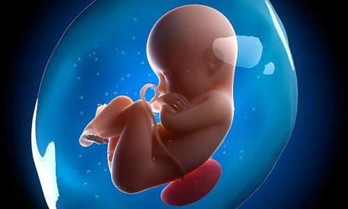
Edwards syndrome is a rare genetic disorder in fetuses. Babies with this condition often have a very low chance of survival after birth, and those who do survive typically experience severe developmental abnormalities. It is the second most common trisomy disorder after Down syndrome, occurring in approximately 1 in 3,000 to 1 in 8,000 newborns.
1. What is Edwards syndrome?
Edwards syndrome, also known as Trisomy 18, is caused by an error in cell division, resulting in an extra chromosome 18 in the genetic makeup. This excess genetic material leads to severe congenital abnormalities in the fetus.
Unlike Down syndrome, which is also caused by an extra chromosome, Trisomy 18 is more life-threatening, especially in the first months of life. The condition occurs in approximately 1 in 5,000 live births, and studies suggest that female infants are more likely to survive than males. However, many affected fetuses do not survive until birth.
2. Effects of Edwards syndrome on the fetus
Most fetuses with Edwards syndrome experience growth restriction in the womb and often stop developing around the 7th month of pregnancy.
Trisomy 18 affects not only the fetus but also the mother, leading to serious complications during pregnancy, such as: Polyhydramnios (excess amniotic fluid), Oligohydramnios (low amniotic fluid), Single umbilical artery, Intrauterine growth restriction (IUGR), Small placenta, Fetal distress, Weak fetal movements.
For babies who survive birth, severe health complications may include:
- Head and facial abnormalities: Small head, small jaw, low-set ears, choroid plexus cysts in the brain, cleft lip, cleft palate
- Short sternum
- Congenital heart defects: Atrial septal defect (ASD), ventricular septal defect (VSD)
- Aortic stenosis
- Spina bifida
- Myelomeningocele
- Abdominal and internal organ defects: Omphalocele, gastroschisis, esophageal atresia, polycystic or hydronephrotic kidneys, horseshoe kidney, undescended testes
- Hand and foot deformities: Clenched hands, hypoplastic nails, clubbed hands, thickened soles
- Clenched fist
- Abnormal kidney
- Undescended testicles (testicles not descending into the scrotum)
- Brain function impairment: Some nerve cells fail to develop fully, clustering in small groups within the brain. This leads to severe intellectual disability and difficulties with essential functions such as sucking, breathing, and swallowing.

3. How is Edwards syndrome diagnosed?
Although there is no cure for Edwards syndrome, it can be detected early through prenatal screening tests.
Prenatal diagnosis:
Two main types of prenatal tests are recommended:
- Screening tests: These estimate the risk of Trisomy 18. They are non-invasive and painless. However, the disadvantage of this test is that it cannot give an exact conclusion about whether the fetus has Edwards syndrome or not, it can only provide couples with useful information to decide whether to perform diagnostic testing or not.
- Diagnostic tests: These provide a highly accurate diagnosis of Trisomy 18 and other chromosomal abnormalities. Amniocentesis is nearly 100% accurate in detecting Edwards syndrome. However, it is an invasive procedure that carries a risk of miscarriage and other fetal complications.
- Postnatal diagnosis: Newborns suspected of having Trisomy 18 can be diagnosed through physical examination, looking for typical external abnormalities such as clenched hands and a shortened sternum.
- NIPT (Non-Invasive Prenatal Testing): NIPT is considered one of the safest and most accurate prenatal screening methods, helping to detect abnormalities in the number and structure of chromosomes in the fetus during pregnancy. NIPT prenatal screening is a non-invasive test, based on fetal DNA testing in the mother's blood.
Vinmec International Hospital offers non-invasive prenatal testing (NIPT) with superior accuracy and safety compared to traditional methods like amniocentesis or chorionic villus sampling (CVS). NIPT can be performed as early as the 9th week of pregnancy, reducing the need for unnecessary invasive procedures.
Dr. Nguyen Anh Tu has over six years of experience in obstetric ultrasound and specializes in prenatal diagnosis. He has received advanced training in fetal ultrasound and diagnosis from the Fetal Medicine Foundation (FMF) and has expertise in prenatal intervention techniques. Dr. Tu has also participated in numerous international fetal medicine conferences and workshops.
Currently, he is practicing at the Department of Obstetrics and Gynecology, Vinmec Hai Phong International Hospital.
Reference source: Trisomy18.org; Webmd.com
To arrange an appointment, please call HOTLINE or make your reservation directly HERE. You may also download the MyVinmec app to schedule appointments faster and manage your reservations more conveniently.