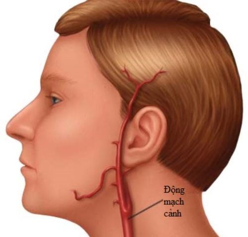
The external carotid artery is one of the two branches of the common carotid artery, supplying blood to the head and neck region. External carotid artery ligation is performed to control bleeding in otorhinolaryngology, maxillofacial, and neurosurgical cases.
1. Indications for surgery
External carotid artery ligation is indicated in the following cases:
- Arterial injury in the head and neck region.
- Persistent nasal bleeding or post-tonsillectomy hemorrhage that does not respond to conventional hemostatic methods, bleeding in oral and tongue cancer.
- Prophylactic ligation in major head and neck surgeries, such as: Maxillary bone resection, Tongue cancer resection, Parotid gland cancer resection, Nasopharyngeal fibroma removal

2. Surgical preparation
Personnel: A Level I otorhinolaryngology specialist with experience in external carotid artery ligation performs the surgery.
An assistant stands opposite the surgeon.
Equipment: 1 standard scalpel, 10 Kocher forceps, 10 Halstead forceps, 1 toothed, and 1 non-toothed dissecting forceps, 1 grooved director, 1 Derchan needle holder, 2 Farabeuf retractors, 1 needle holder and needles, 5 surgical towel clamps, 1 straight scissors, 1 long curved scissors, 1 long (20 cm) Kocher forceps, Sutures
Patient preparation: The patient is urgently prepared and stabilized with intensive resuscitation and blood transfusion.
If the patient has not lost a significant amount of blood, they may receive intramuscular injections of: Dolargan 0.10; Pipolphen 0.05; Atropine 1/4 mg x 2 ampules
Medical records: Complete medical records, including basic lab tests and lateral neck X-rays. Ultrasound imaging if available.
3. Surgical procedure
1% Xylocaine is injected along the anterior border of the sternocleidomastoid muscle over a 10 cm length. The patient lies in a supine position with a pillow under the shoulders and the head turned to the opposite side. Antiseptic solution is applied, and sterile drapes are placed.
Step 1: Skin and superficial fascia incision
- An 8 cm incision is made starting at the level of the mandibular angle, 1 cm posterior to it.
- The incision follows the anterior border of the sternocleidomastoid muscle down to the level of the cricoid cartilage.
- The first cut goes through the skin, subcutaneous tissue, platysma muscle, and fat.
- The second cut divides the superficial cervical fascia along the anterior border of the sternocleidomastoid muscle.
- If the external jugular vein is encountered, clamped and ligated.
Step 2: Identifying the Farabeuf ttriangle
- The sternocleidomastoid muscle is retracted laterally, and careful dissection is performed in the carotid sheath.
- The Farabeuf triangle is defined by the internal jugular vein, the common venous trunk (linguofacial vein), and the hypoglossal nerve (running transversely below the digastric muscle belly).
Step 3: Identifying the external carotid rtery using two criteria
- The external carotid artery is medial, while the internal carotid artery is lateral.
- The external carotid artery gives off branches, whereas the internal carotid artery does not. (Identification should be done within the Farabeuf triangle.)
Step 4: Ligation of the external carotid artery
- Novocaine is injected into the carotid sheath at the bifurcation of the internal and external carotid arteries to suppress carotid body reflex stimulation.
- A dissecting forceps is used to pass a suture around the artery, and vicryl or perlon sutures are placed.
- The ligation site should be 1–2 cm above the bifurcation of the common carotid artery into internal and external branches (not directly at the bifurcation).
- Selective ligation of individual branches (thyroid, lingual, or facial artery) may be performed if necessary.
Step 5: Closure: Small vessels are ligated using catgut. The incision is closed in two layers: The inner layer with catgut, the outer layer with linen or nylon sutures.
4. Postoperative monitoring and complication management
Dressings are changed every three days. Sutures are removed after one week.
Rare complications
Unilateral ligation:
- Cerebral embolism: Caused by dislodged thrombi entering the internal carotid artery and traveling to the brain.
- Severe carotid sinus reflex: May result in dizziness, ipsilateral hearing loss, and facial pallor. It can be managed with xylocaine injections but can sometimes be fatal.
- Neurological complications: Hemiplegia, speech impairment.
- Bilateral Ligation: Typically uneventful but may lead to pallor and, in rare cases, death within a few hours.
Timely external carotid artery ligation prevents blood flow to its branches, effectively controlling hemorrhage in its dependent regions. This improves treatment outcomes and enhances patient recovery.
SEE MORE
- How dangerous is carotid stenting?
- Doppler ultrasound of the carotid artery
- Carotid stenosis: Causes, symptoms, diagnosis and treatment
To arrange an appointment, please call HOTLINE or make your reservation directly HERE. You may also download the MyVinmec app to schedule appointments faster and manage your reservations more conveniently.