Article written by Mai Viễn Phương, MSc, MD, Department of Outpatient & Internal Medicine - Vinmec Central Park International Hospital
Gastroesophageal reflux disease (GERD) is a significant economic burden that greatly reduces the quality of life and work productivity of patients, and its prevalence is increasing in Asian countries, especially Vietnam. The incidence of gastroesophageal reflux disease (GERD) in Vietnam has recently reached 15.4% among patients with upper gastrointestinal symptoms, which is higher than the prevalence of gastric ulcers (8.2%) and duodenal ulcers (6.7%). The Hill grading of the gastroesophageal junction (GEJ) has been shown to be a simple and useful tool for predicting gastroesophageal reflux, even more effective than lower esophageal sphincter pressure.
Where is the stomach located in the body?
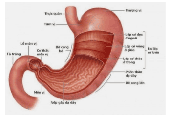
The stomach is the widest part of the digestive tract, connecting above with the esophagus and below with the duodenum. Structurally, the stomach has four main parts: the cardia, fundus, body, and pylorus. The cardia is the junction where the esophagus meets the stomach, and food from the esophagus passes through the cardia into the stomach without a closing valve, only having mucosal folds known as the gastroesophageal folds. The fundus is dome-shaped, located below the diaphragm, above, and to the left of the cardia. Underneath the fundus is the body, which is the main part of the stomach. The pylorus is funnel-shaped and connects the stomach to the duodenum. The pyloric sphincter (a type of smooth muscle) is located at the end of the junction between the stomach and the duodenum.
Where is the gastroesophageal fold located?
The gastroesophageal fold is located at the junction between the esophagus and the stomach.
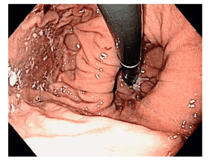
During digestive endoscopy, when examining various parts of the stomach in an upright position, the endoscopist will maneuver the scope to investigate the regions of the fundus and cardia, observing the mucosal folds converging at the junction of the esophagus and stomach, which is the gastroesophageal fold.
What is gastroesophageal reflux disease (GERD)?
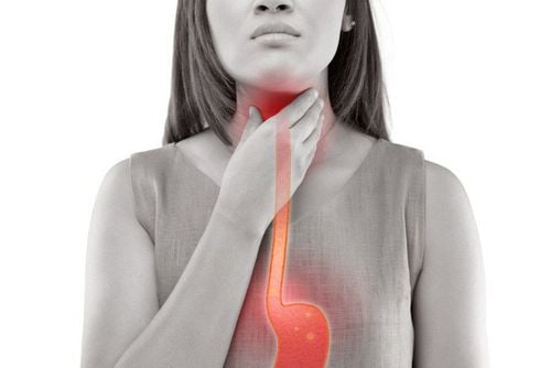
Esophagitis is the inflammation of the mucosal lining inside the esophagus, the tube connecting the throat to the stomach that serves the function of delivering food into the body. The condition can be caused by acid reflux, infections, side effects of certain medications, or food allergies, leading to difficulties in swallowing and chest pain.
Mechanism of gastroesophageal reflux disease
Gastroesophageal reflux disease occurs when stomach acid regularly flows back into the esophagus. This acidic fluid can irritate the lining of the esophagus.
Relationship Between Gastroesophageal Reflux Disease (GERD) and the Gastroesophageal Junction

Gastroesophageal reflux disease (GERD) is a common disorder with a prevalence of about 10-20% in Western countries. The clinical manifestations of GERD (i.e., acid regurgitation and heartburn) are due to the backflow of digestive juices from the stomach into the esophagus. The current understanding of the pathogenesis of GERD is multifactorial, involving the lower esophageal sphincter (LES), diaphragm, esophageal acid neutralization, gastric acid secretion, gastric emptying, and intra-abdominal pressure. The Montreal definition of gastroesophageal reflux disease states that GERD occurs when the backflow of stomach contents into the esophagus causes symptoms and/or complications. Possible complications include esophagitis and Barrett's esophagus. In Barrett's esophagus, the normal squamous epithelium of the distal esophagus is replaced by columnar epithelium, leading to the development of intestinal-type columnar epithelium in the esophagus. The definition of Barrett's esophagus is often debated, and there is no universally accepted criteria. The most common definition of Barrett's esophagus requires histologically verified intestinal metaplasia at the gastroesophageal junction.
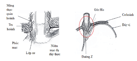
The gastroesophageal junction (GEJ) is the anatomical area where the distal esophagus connects to the proximal stomach. Under normal conditions, it is at the level of the diaphragm. However, the position of the GEJ is not fixed and can move several centimeters during swallowing and breathing. During swallowing, the longitudinal smooth muscle of the esophagus contracts, shortening the esophagus, leading to a physiological hernia. The gastroesophageal junction (GEJ) is then returned to its original position by elastic support structures, particularly by the esophageal-diaphragmatic membrane. When the GEJ, along with the lower esophageal sphincter (LES) and the stomach cardia, is extended upward into the thoracic cavity through the diaphragm, a hiatal hernia will appear. Studies have shown that hiatal hernia reduces LES pressure and the sphincter function of the diaphragm. The presence and axial length of the interrupted hernia mass have also been shown to correlate with the severity of gastroesophageal reflux disease (GERD).
Evaluation of the Hill grading system for abnormalities of the gastroesophageal flap valve (GEFV):
The characteristics of the gastroesophageal flap valve (GEFV) according to the Hill classification are documented to be related to gastroesophageal reflux disease (GERD). In Vietnam, the prevalence of GERD tends to be increasingly common, but there are currently very few studies on the GEFV.
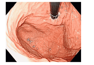
The characteristics of the GEFV according to the Hill grading system are evaluated as shown in Figure 1. (A) Grade I: clear folds, the cardia tightly embraces the endoscope. (B) Grade II: folds are still clear but not as clear as grade I, the cardia opens and closes quickly with breathing. (C) Grade III: folds are not clear, and the cardia does not tightly embrace the endoscope. (D) Grade IV: no folds are present, the gastric-esophageal junction is open, and the esophageal epithelium can be seen in the retroflexed position of the endoscope in the stomach.
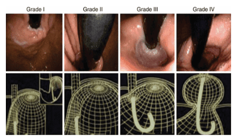
(A) Grade I: clear folds, the cardia tightly embraces the endoscope. (B) Grade II: folds are still clear but not as pronounced as grade I, the cardia opens and closes quickly with breathing. (C) Grade III: folds are not clear, and the cardia does not tightly embrace the endoscope. (D) Grade IV: no folds are present, the gastric-esophageal junction is open, and the esophageal epithelium can be seen in the retroflexed position of the endoscope in the stomach.
Are abnormalities of the gastroesophageal flap valve common?
Due to the increasing incidence of GERD in Asia, many research studies have been conducted to explore risk factors for the disease in Asia. Known risk factors include age, male gender, smoking, and obesity. The abnormality of the GEFV has also been mentioned in some studies, indicating that GEFV abnormality is an independent factor related to GERD. There is very little data from Asia on the prevalence of GEFV abnormalities. Iwamoto and colleagues reported an abnormal GEFV prevalence in Japan of 13.5%, with a GERD prevalence of 27% among patients undergoing upper gastrointestinal endoscopy. In another study in Taiwan, Lin and colleagues reported a GEFV abnormality prevalence of 27.3%, with a GERD prevalence of 41.3% among healthy individuals undergoing gastric cancer screening endoscopy. According to studies by several authors in Vietnam, the prevalence of GEFV abnormalities was found to be 36.2%, with the majority being grade III and very few cases in grade IV. Similar to previous studies in other populations, the authors also found that GEFV abnormalities are an independent risk factor for GERD. This result helps explain why the prevalence of GERD tends to be higher in populations with a higher prevalence of GEFV abnormalities. A recent study comparing two groups of patients: Caucasian Russians and Koreans also found that GEFV abnormalities are an independent risk factor for GERD regardless of ethnicity. According to many studies, the GEFV abnormality is more common in males compared to females and more frequently in individuals under 40 years of age compared to those over 40. Contractor and Iwamoto's research indicates that the rate of abnormal flap valve increases with age, while other studies have not found a correlation between age and flap valve abnormalities. This correlation lacks a satisfactory explanation and requires further investigation in the future.
Is there a relationship between GEFV abnormalities and obesity?
Many authors’ studies have not found a relationship between GEFV abnormalities and obesity, abdominal obesity, smoking, or alcohol consumption.
The role of esophagogastroduodenoscopy in assessing abnormalities of the gastroesophageal flap valve.
Esophagogastroduodenoscopy (EGD) is the standard method for evaluating the upper gastrointestinal tract. The ability of the mechanical anti-reflux barrier can be endoscopically assessed in two ways: first, measuring the axial length of any hiatal hernia present (between the hernia and the gastric-esophageal junction). Due to the physiological dynamics in this area, it can be difficult to measure the length of the hiatal hernia, even under ideal conditions. It is unclear at what length a hiatal hernia becomes clinically significant, meaning pathological significance, and since the GEJ is not fixed, most endoscopists use a length of 2 cm as the diagnostic threshold between normal and abnormal. Another way to assess the GEJ is to classify the gastroesophageal flap valve (GEFV) using the Hill classification. Studies have shown a correlation between the Hill grading system and the prevalence of GERD. Hill classification at a high level is also related to low lower esophageal sphincter pressure, increased incidence of hiatal hernia, and has the ability to predict poor response to proton pump inhibitor therapy. The Hill classification has been shown to be appropriate, useful, and provides useful information when assessing patients suspected of having GERD. Esophagitis can be identified through endoscopy and classified according to the Los Angeles (LA) classification.
In addition to known risk factors for reflux esophagitis such as overweight or obesity, smoking, alcohol consumption, eating fried or spicy foods, and eating just before sleeping, abnormalities of the gastroesophageal junction valve have also been recorded as an independent risk factor for this condition. At the same time, abnormalities of the valve also help predict the treatment response of the patient.
Vinmec International General Hospital is a reputable address trusted by many patients in performing diagnostic and treatment techniques for esophageal-stomach, rectal-colon diseases, etc. Additionally, at Vinmec Hospital, colonoscopy diagnostics are conducted using the Olympus CV 190 endoscope, featuring NBI (Narrow Band Imaging), which provides clearer mucosal pathology analysis images compared to conventional endoscopy, enabling early detection of abnormal mucosal lesions. Vinmec Hospital, with modern facilities and equipment along with a team of experienced specialists, is always dedicated to patient care; clients can be assured of digestive endoscopy services at Vinmec International General Hospital.
Customers interested in digestive disease diagnostic services at Vinmec and needing advice and support can book an online consultation on the website or contact Vinmec's hotline system for detailed advice.
To arrange an appointment, please call HOTLINE or make your reservation directly HERE. You may also download the MyVinmec app to schedule appointments faster and manage your reservations more conveniently.