Currently, the incidence of kidney and urinary tract-related diseases is very common. Renal ultrasound is one of the effective diagnostic imaging methods for detecting kidney enlargement. Based on the enlarged kidney images displayed on the ultrasound results, doctors can diagnose many serious conditions affecting the kidneys.
The article is professionally consulted by Nguyen Van Huong, MSc, MD - Department of Diagnostic Imaging - Vinmec Da Nang International General Hospital.
1. Principles of Kidney Function
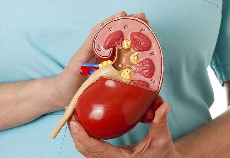
In the body, the kidneys are vital organs of the urinary system, with each person having two kidneys symmetrically located on either side of the lumbar spine. The kidneys function to filter waste and convert it into urine, maintain acid-base balance, regulate electrolytes, and control blood pressure.
The kidneys act as the body's natural blood filter. It is estimated that each day, 8 liters of blood are filtered through the kidneys 20-25 times, corresponding to these organs filtering about 180 liters/24 hours. The components in the blood continuously change as we digest food and drinks, meaning the kidneys must work continuously without rest.

2. What is Renal Ultrasound?
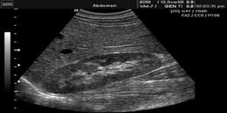
Ultrasound is a diagnostic technique that uses sound waves to generate images of the size, structure, and pathological signs of various organs and tissues in the body, including the kidneys. Renal ultrasound is particularly useful in diagnosing kidney conditions such as kidney stones, kidney cysts, kidney abscesses, and hydronephrosis. It is also a non-invasive, painless method with high accuracy and safety for patients.
Doctors often recommend renal ultrasound for patients with symptoms such as difficulty urinating, urinary retention, hematuria, kidney and ureteral trauma and pain, acute and chronic kidney failure, absence of kidney shadow on X-ray, sudden hypertension, or suspected polycystic kidney disease.
3. Signs and Causes of Kidney Enlargement
3.1 Normal Kidneys on Ultrasound
The kidneys have an oval shape, with the renal hilum located on the inner side. In adults, the right kidney tends to be lower than the left kidney. The average size of the kidneys is 9-12cm in length, 4-8cm in width, and 3-5cm in thickness. If the kidney size exceeds these measurements, it is considered kidney enlargement. The size of the two kidneys can differ by 1-1.5cm and can vary based on gender (men have larger kidneys than women) and age. On a healthy kidney ultrasound, the borders appear smooth, the left kidney is compressed by the spleen, making the renal parenchyma appear triangular, and the renal arteries and veins are visible.

3.2 Causes of Kidney Enlargement
Conversely, an enlarged kidney on ultrasound indicates a pathological condition of the kidney. The causes of kidney enlargement can be due to two reasons:
- Hyperplasia of kidney cells causing kidney enlargement (e.g., kidney cancer...)
- Hydronephrosis due to obstruction below, such as ureteral stones, ureteral stricture…
Specifically, kidney enlargement can be due to conditions such as:
- Hydronephrosis, pyonephrosis:
Due to some obstruction in the urinary tract, urine accumulates in the renal pelvis, causing kidney enlargement. Prolonged urine retention allows bacteria to develop, gradually turning into pus, causing fever, kidney swelling and pain, and pus in the urine.
Causes of hydronephrosis and pyonephrosis include:
- Kidney stones, ureteral stones.
- Abdominal tumors pressing on the ureter or a branch of the iliac artery crossing the ureter…
- Renal tuberculosis causes ureteral stricture, leading to ureteral and renal pelvis stricture.
- Prostate tumors, brain injuries, spinal cord inflammation causing urinary retention, and urinary obstruction.
Polycystic kidney disease:
A congenital malformation resulting in an ultrasound image of enlarged kidneys (usually on both sides, with a lobulated surface). Patients often experience lumbar pain, sometimes renal colic, and if there is bacterial superinfection, fever or cloudy urine (urine contains protein, many red blood cells, white blood cells), and chronic high blood urea. Ultrasound images also show indirect images of "renal cysts" and elongated renal calyces. Polycystic kidney disease progresses slowly, potentially lasting many years, but patients may still die from bacterial superinfection, kidney failure, and high blood urea.
Kidney cancer:
A common malignancy in the elderly, more frequent in men than women. Ultrasound of kidney cancer patients shows enlarged, hard kidneys with a rough surface, a mass occupying the kidney, and may be associated with varicocele. Patients experience lumbar pain, hematuria, and urine containing many red blood cells.
Compensatory kidney enlargement:
If the body has only one kidney, the remaining kidney must work harder, causing it to enlarge (sometimes up to 1.5 times normal size). This compensatory kidney enlargement is not pathological, and urine tests will not show protein, white blood cells, red blood cells, and blood urea and creatinine levels are not high.
Compensatory hypertrophy:
Similar to compensatory kidney enlargement, occurring when the opposite kidney is absent, non-functional, or has reduced function. The working kidney increases in length and cross-sectional area by up to 30%, with volume increasing by up to 80%. The morphology of the kidney remains normal.
- Acute pyelonephritis: This condition can cause kidney enlargement in both length and cross-sectional area. However, there are cases where acute pyelonephritis shows very little or no change in size.
- Renal vein thrombosis: This condition also causes kidney enlargement. Ultrasound shows changes in kidney echogenicity with alternating hyperechoic and hypoechoic areas due to bleeding and swelling, and perirenal fluid collections may appear.
- Acute arterial infarction: This rare condition can also cause kidney enlargement. Hyperechoic and hypoechoic areas of edema and bleeding are similar to those seen in renal vein thrombosis.
- Infiltrative causes: Amyloidosis, lymphoma, and many other infiltrative causes also lead to kidney enlargement, with loss of corticomedullary differentiation. There is no distinction between different infiltrative causes.
- Acute tubular necrosis: This condition often causes kidney enlargement in severe cases. Kidney size changes more in cross-sectional area than in length. The kidneys may appear normal if the cause is ischemia, but will show increased cortical echogenicity if nephrotoxic, and the renal pyramids will enlarge with signs of Tamm Horsfall protein.
Nguyen Van Huong, MSc, MD, was a lecturer in the Department of Medical Imaging at Da Nang University of Medical Technology and Pharmacy, with many years of experience working at hospitals such as: Cancer Hospital; Da Nang Hospital; Da Nang Obstetrics and Pediatrics Hospital.
With his dedication to his work and his significant contributions to the medical field and the diagnostic imaging specialty in particular, Dr. Huong is always loved and trusted by patients.
To arrange an appointment, please call HOTLINE or make your reservation directly HERE. You may also download the MyVinmec app to schedule appointments faster and manage your reservations more conveniently.


