Learn about lower extremity computed tomography
This is an automatically translated article.
The article was professionally consulted by Specialist Doctor I Tran Cong Trinh - Radiologist - Radiology Department - Vinmec Central Park International General Hospital.
1. What is lower extremity artery disease?
The main cause of lower extremity artery disease is atherosclerosis. Risk factors for atherosclerosis include:
Age (common in ages 55-60); gender (male/female = 3/1); Cigarette; Diabetes mellitus; hypertension; Dyslipidemia; Increased homocysteine levels in the blood.
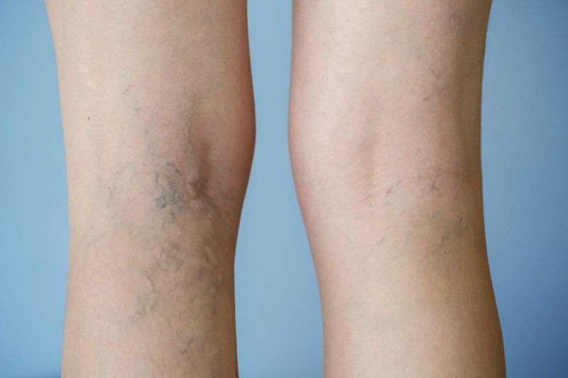
2. Varicose veins of the lower extremities

3. What is lower extremity computed tomography?
Computed tomography of the lower extremities is often indicated for the next generation of multi-slice tomography, preferably 64 sequences or more because of the requirement for fast cutting to keep up with the hemodynamics of iodinated contrast agents. blood vessel lumen.
3.1. Indications for computed tomography arterial angiography of the lower extremities in patients with acute and chronic arterial occlusion. Patients with aneurysms, vascular malformations. Check after stent placement. Evaluation of normal anatomy and abnormality of lower extremity arterial system 3.2. Contraindications to lower extremity computed tomography angiography There are no absolute contraindications. Contraindications for intravenous iodine contrast agents, patients with a history of allergic disease such as bronchial asthma, liver failure, kidney failure, especially in patients with a history of allergy to iodinated contrast agent.
4. Preparing for lower extremity computed tomography

5. Steps to perform lower extremity computed tomography
5.5 Evaluation of results Image results show anatomical vascular structures in the examination area Detect lesions if any Conclusion: In the techniques related to lower extremity angiography, microscopic tomography Lower extremity arteriosclerosis is an accurate diagnostic imaging procedure that effectively aids in the detection of lower extremity vascular diseases.
Any questions that need to be answered by a specialist doctor as well as customers wishing to be examined and treated at Vinmec International General Hospital, you can contact Vinmec Health System nationwide or register online HERE.
MORE:
Computed tomography - the gold standard in diagnosing coronary heart disease Coronary artery disease: Symptoms, causes and treatment in Vinmec Intravascular Ultrasound (IVUS) - An effective "assistant" help in interventional treatment of coronary artery disease
-
 Laser treatment of varicose veins of the lower extremities
Varicose veins of the lower extremities progress silently, can cause complications such as superficial vein thrombosis and deep vein thrombosis, causing pain and edema of the lower extremities. Pathological causes may be related to genetic factors, occupations requiring long standing, ...
Readmore -
 Symptoms of varicose veins
Because the disease progresses silently, recognizing the signs of varicose veins is extremely important for patients to go to the hospital for examination and timely treatment.
Readmore -
 How to improve lower extremity venous system function?
Hi doctor. I have varicose veins in my lower extremities, often have numbness and pain in my legs at night, standing for a long time feel very heavy. When it turns to heaven, the legs are numb and restless, unable ...
Readmore -
 Development and treatment of varicose veins of the lower extremities
Venous insufficiency of the lower extremities worldwide accounts for a significant proportion, of which women account for 70%. Although the disease can be less dangerous, it can cause tight calves, heaviness and fatigue in the legs, night cramps, crawling sensations, ...
Readmore -
 Treatment of varicose veins in mothers after childbirth
Giãn tĩnh mạch là những tĩnh mạch lớn so với bình thường, sưng thường xuất hiện ở chân và bàn chân. Chúng xảy ra khi các van trong tĩnh mạch không hoạt động như bình thường, do đó máu không ...
Readmore