Beware of mammary gland calcifications
This is an automatically translated article.
1. What is calcification?
When a specific cause cannot be found, the description of calcifications should include both their morphology and distribution. It is not necessary to always report obvious benign calcifications. However, they should be reported if the physician reading the film is concerned that other film readers may misinterpret them. Vascular calcification was noted in the results, particularly in women younger than 50 years of age for whom data suggest a potential risk of coronary artery disease.

2. What is mammary gland calcification?
Mammary calcification can be defined as the deposition of calcium salts in the female breast tissues. Calcium deposits can be so small that you can't feel them yourself and cause no pain. In general, mammary gland calcifications are usually benign and not much of a concern. However, in certain cases, breast calcifications can be an early sign of breast cancer.
There are 2 types of mammary calcifications that can be detected by mammography:
Large calcifications that appear as large round and white spots on a mammary gland and are randomly dispersed in the breast tissue. Large calcifications are quite common in women over the age of 50, which is a natural sign of breast aging; Microcalcifications or small calcified nodules are very small in size, appearing with many small white spots on mammograms. However, if they appear in clusters or are not limited, small calcified nodules can be a sign of cancer, which needs to be fully examined and diagnosed by a specialist.
3. Causes of mammary gland calcification
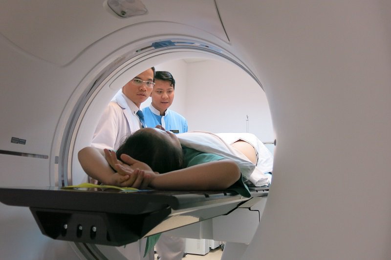
Additional tests may include mammogram with magnification of the calcified areas of the mammary gland, or even biopsy to determine the nature of the calcium deposits, images used to Comparison of calcium deposition patterns in breast tissues.
4. Treatment of calcified mammary glands
Procedural sampling: The doctor will rely on the results of the mammogram and ultrasound to perform this procedure and mark it. Then, a small piece of breast tissue is removed from that area under general anesthesia in a hospital operating room. Finally, the biopsy sample is sent for examination in the pathology laboratory; Needle core biopsy: This procedure is also done frequently. The surgeon cuts out small pieces of tissue with a small, hollow needle with the help of a computer-guided device. The patient is under local anesthesia and does not require surgery, then the removed tissue sample is sent to the pathology laboratory for examination. Breast biopsy with vacuum-assisted suction under Stereotactic positioning: This technique uses a large biopsy needle with a size of 12 G or more, combined with vacuum suction, so it helps to get more specimens, thereby increasing the probability of success. In addition, this technique is also less invasive than the open biopsy method: The incision site does not require any recovery stitches, does not require special care, and the patient can return to normal daily life right away. tomorrow. In addition, the placement of markers to mark the intervention site is carried out right in the procedure to help manage the lesions after the test results are more favorable. This is the most modern technique today, being deployed by Vinmec Times City (Hanoi) and very few hospitals in Vietnam.
Please dial HOTLINE for more information or register for an appointment HERE. Download MyVinmec app to make appointments faster and to manage your bookings easily.
-
 Fine-needle aspiration technique for palpable breast lesions
Sử dụng phương pháp chọc hút bằng kim nhỏ trong các tổn thương vú có thể sờ nắn được sẽ giúp các bác sĩ chẩn đoán chính xác tình trạng khối u ác hay lành tính và nguyên nhân hình ...
Readmore -
 In which cases is a breast biopsy under X-ray guidance indicated?
Sinh thiết vú dưới hướng dẫn X-quang là kỹ thuật dùng kim sinh thiết kích thước lớn gắn với máy hút, sử dụng máy X-quang làm hướng dẫn để điều khiển kim vào đúng vị trí nhằm lấy các mảng ...
Readmore -
 Surprising things that could help treat breast cancer
Breast cancer is a disease with a high mortality rate. However, if breast cancer is detected and treated effectively, the patient still has a chance to prolong life. Caring for cancer patients with appropriate methods will bring high results for ...
Readmore -
 In what situations does aortic stenosis occur?
Aortic valve stenosis is a dangerous heart valve disease with a high incidence, especially in the elderly. What causes aortic valve stenosis? How to vet? The following article will help you better understand this disease.
Readmore -
 Elderly people with diabetes, high blood pressure can do biopsies?
Hello doctor, my mother is 71 years old, found to have a lump in her left breast. The doctor ordered a biopsy. My mother has diabetes and high blood pressure, can I have a biopsy? Looking forward to consulting your ...
Readmore