The article has been consulted by Dang Manh Cuong, M.A., M.D. - Diagnostic Imaging Physician - Department of Diagnostic Imaging - Vinmec Central Park International General Hospital. The doctor has over 18 years of experience in ultrasound - diagnostic imaging.
Ultrasound of the axillary lymph node is a simple imaging diagnostic method that helps doctors diagnose certain diseases of the axillary lymph nodes. This method is also the first-line approach in cases of abnormalities in the axillary lymph nodes and suspected metastatic lesions in cancer.
1. What is axillary lymph node ultrasound?
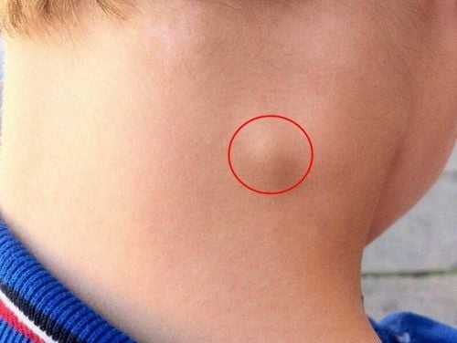
The axillary lymph node is one of the lymph nodes in the body, involved in the process of eliminating or destroying harmful microorganisms. Normally, the axillary lymph node is not palpable; however, in some cases, the lymph node becomes swollen due to the body’s immune response, making it detectable. The enlargement of lymph nodes can be caused by various factors, and to help identify the underlying cause of axillary lymph node enlargement, ultrasound imaging is used.
Axillary ultrasound is a non-invasive, painless technique that uses a high-frequency linear ultrasound probe to examine the axillary region and detect abnormal lymph nodes or pathological lesions of lymph nodes. Based on the images obtained during the ultrasound, doctors can provide directions for diagnostics.
Sometimes, lymph node enlargement occurs as a normal response after receiving the tuberculosis vaccine. In this case, the ultrasound may reveal a solitary lymph node, typically round or oval in shape, hypoechoic compared to surrounding tissue, with smooth borders, and possibly fluid-filled,...
In addition to common inflammatory reactions, swollen lymph nodes may also indicate abnormalities that need further diagnosis. With axillary lymph node ultrasound, conditions such as metastatic cancer or tuberculous lymphadenopathy can be detected through suggestive signs on the ultrasound images.

2. Indications and contraindications for axillary lymph node ultrasound
Indications for axillary lymph node ultrasound:
- Suspected cancerous lesions in organs that may metastasize to the axillary lymph nodes, such as breast cancer, lymphoma, stomach cancer, …
- Abnormal swelling or pain of lymph nodes.
- Ultrasound-guided lymph node biopsy or fine-needle aspiration (FNA) for diagnosing whether the lymph node lesion is benign or malignant.
Contraindications: This is a non-invasive procedure with no contraindications unless accompanied by additional interventions. In cases where ultrasound is used to guide biopsy or FNA, contraindications include blood clotting disorders.
3. Steps for Performing Axillary Lymph Node Ultrasound
3.1 Preparation:
- The procedure is performed by a diagnostic imaging physician, ultrasound technician.
- Equipment: A flat probe ultrasound machine with a frequency of over 7.5 MHz, ultrasound gel, and cloth to wipe off the gel after the procedure.
- Patient: No special preparation. The patient is clearly explained about the diagnostic ultrasound procedure to cooperate during the examination.

3.2 Procedure steps
Patient position: lie on their back, with the arm on the side to be examined raised above the head or placed under the patient's neck to expose the axillary area that needs to be ultrasounded.
Procedure steps:
- Step 1: Perform ultrasound on both breasts first to detect any abnormalities in all regions, from the areola outward to the periphery. Typically, the ultrasound probe is moved in a circular motion, either clockwise or counterclockwise, starting from the center of the breast to the outermost area.
- Step 2: The probe is placed to scan the axillary region, with cross-sectional cuts perpendicular to the head of the humerus, allowing clear visualization of the glenohumeral joint on the examined side and the joint space, as well as the bony contours of the humerus and scapula. Any lesions in this area should be examined.
- Step 3: Identify the brachial blood vessels on the transverse section, including the artery and vein. It is best to monitor the blood vessels using color Doppler ultrasound.
- Step 4: Examine the lymphatic system by rotating the probe along the vascular bundle, moving downward parallel to the bundle towards the anterior axillary line. If multiple lymph nodes with lesions are found at different locations, create a diagram marking their position, depth, and distance from the skin and major blood vessels for easier guidance for cytological diagnosis or histopathological diagnosis.
- Step 5: The results should describe the location, size, shape, borders, capsule, number, and blood flow of the lesion, along with other necessary tests for a diagnosis. This will help the clinical doctor determine the diagnosis and further recommendations:
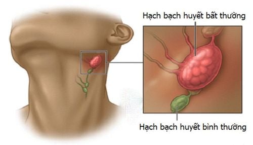
- Normal lymph node: Small in size, usually a few millimeters, with a thin capsule, and an eccentric hilum with well-defined borders.
- Reactive inflammatory lymph node: Often presents as multiple discrete nodes, small and similar size, hypoechoic, well-defined, oval or flat shape, with a hilum, and possibly increased central blood flow…
- Malignant lymph node: Typically, large over 3 cm in size, with a thick capsule, heterogeneous echogenicity, unclear borders, calcifications within the node, and increased peripheral blood flow… In cases suspected of malignancy, doctor will prescribe further tests such as biopsy to confirm the diagnosis.
If ultrasound is performed without a diagnostic needle aspiration, no special follow-up is required after the procedure.
Axillary lymph node ultrasound is a simple, inexpensive, and quick method for detecting certain lymphatic diseases. It also helps guide the diagnosis of some malignant diseases, enabling early detection if abnormalities occur.
To arrange an appointment, please call HOTLINE or make your reservation directly HERE. You may also download the MyVinmec app to schedule appointments faster and manage your reservations more conveniently.