This article was professionally consulted by an Obstetrician and Gynecologist - Department of Obstetrics and Gynecology - Vinmec Phu Quoc International General Hospital
A normal fetal umbilical cord contains two umbilical arteries and one umbilical vein encased in Wharton’s jelly. A single umbilical artery is a common umbilical cord abnormality that can pose risks to the fetus's health if not detected early.
1. What is a single umbilical artery?
A normal umbilical cord includes two umbilical arteries and one umbilical vein, surrounded by Wharton’s jelly to nourish and protect the blood vessels. The umbilical vein carries oxygen-rich blood from the mother through the placenta to nourish the fetus, while the umbilical arteries return oxygen-poor blood from the fetus to the placenta for metabolic exchange. The umbilical vein has a larger diameter and is located deeper than the arteries.
A single umbilical artery occurs in approximately 1% of pregnancies, where the cord contains only two blood vessels. It is more frequently found in multiple pregnancies compared to singleton pregnancies. This anomaly may appear in isolation or alongside defects in other systems, such as the cardiovascular, nervous, urinary, or skeletal systems, or as an indicator of chromosomal abnormalities. When isolated, a single umbilical artery generally has a good prognosis, and the fetus's health and development are considered normal. With advancements in diagnostic imaging, such as ultrasound, single umbilical artery can be detected prenatally, often along with other congenital defects if present. Definitive diagnosis is confirmed post-delivery through examination.

2. Causes of single umbilical artery
The exact cause of a single umbilical artery remains unclear. Several theories explain its occurrence:
- Failure of one umbilical artery to develop early in pregnancy.
- Regression of one umbilical artery during pregnancy development. In this case, the umbilical cord initially has three blood vessels but one artery disappears later.
- It can be a standalone abnormality or associated with chromosomal abnormalities such as Down syndrome or Edwards syndrome.
Risk factors for a pregnancy with a single umbilical artery include:
- Maternal age over 40.
- A history of multiple previous births.
- Multiple pregnancies.
- Female fetus.
- Maternal smoking.
- Maternal medical conditions such as diabetes or hypertension.
- A single umbilical artery can also occur in pregnancies without these risk factors.
Therefore, regular prenatal check-ups and ultrasound screenings for fetal anomalies are essential for a healthy pregnancy.

3. Diagnosis of single umbilical artery
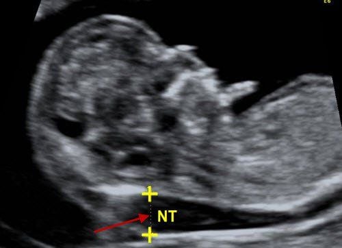
Ultrasound is the primary tool for diagnosing a single umbilical artery and detecting accompanying abnormalities. This imaging technique is non-invasive, safe for both mother and fetus, and can be performed multiple times. Approximately one-quarter of cases with a single umbilical artery may lead to complications such as preterm labor, preterm birth, or intrauterine growth restriction. In some cases, it may be associated with anomalies in other organ systems, such as the nervous, cardiovascular, urinary, or skeletal systems. Prenatal ultrasound can identify associated anomalies, guiding decisions on further interventions, such as amniocentesis. Regular fetal growth monitoring through serial ultrasounds can detect developmental delays early, allowing timely management.
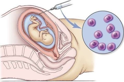
Additional diagnostic methods, such as non-invasive prenatal testing, amniocentesis, or chorionic villus sampling, may also be applied in diagnosing and managing single umbilical artery abnormalities, with these tests being guided by prior ultrasound findings.
4. Principles of delivery management in pregnancies with single umbilical artery
A single umbilical artery has a good prognosis if it is isolated and not accompanied by other congenital defects. Pregnant women diagnosed with this condition should receive proper counseling on prognosis and appropriate management strategies. During labor, the following principles should be followed:
- A single umbilical artery is not an indication for cesarean delivery.
- Vaginal delivery can proceed as in a normal pregnancy.
- Fetal heart monitoring should be closely observed.
- Postnatal evaluation of the cardiovascular and urinary systems is recommended.
- Breastfeeding should be encouraged.
- A single umbilical artery is a common umbilical cord abnormality that can pose risks to fetal health if undetected. Therefore, expectant mothers should attend regular prenatal check-ups and not skip screenings for fetal anomalies.

Realizing the importance of screening for fetal abnormalities, Vinmec International General Hospital has and continues to deploy a package maternity service, which was born as a solution. The method helps pregnant mothers feel secure because they have the companionship of the medical team throughout the pregnancy from care, monitoring, comprehensive examination, ultrasound testing and health advice for pregnant women to healthy developing fetus. Pregnant women can rest assured and relax during pregnancy, without having to worry about missing appointments and making appointments. In particular, maternity packages also come with many gift programs and free prenatal classes. When going to give birth, a pregnant mother does not need to prepare too many things because Vinmec has already prepared essential items for mother and child during childbirth and convalescence at the hospital.
Currently, to improve service quality, Vinmec is also equipped with a system of ultrasound machines and modern medical equipment. Accordingly, the examination and diagnosis process at Vinmec is carried out by qualified and well-trained doctors, so it will soon detect obstetric problems and pathologies, fetal malformations from early (if any) for timely examination and treatment right after the baby is born.
To arrange an appointment, please call HOTLINE You may also download the HERE. Download MyVinmec app to schedule appointments faster and manage your reservations more conveniently.
To arrange an appointment, please call HOTLINE or make your reservation directly HERE. You may also download the MyVinmec app to schedule appointments faster and manage your reservations more conveniently.