The CD3, CD4, CD8 cell count test is a blood test used to assess immune system activity in individuals with immunodeficiency. This test evaluates cell surface markers for immune cells. It is widely applied and provides reliable results.
1. What are CD3, CD4, and CD8 cells?
- CD3 is a common T lymphocyte antigen found on the surface of all T lymphocytes.
- CD4 is an antigen on the surface of helper T lymphocytes (T-helper cells), monocytes, and some activated granulocytes.
- CD8 is an antigen found on the surface of T lymphocytes. It is toxic and suppressive to T lymphocytes.
2. What is the CD3, CD4, CD8 cell count test?
The CD3, CD4, and CD8 cell count test
uses flow cytometry techniques that use monoclonal antibodies against CD3, CD4, and CD8 with different fluorescent colors. This enables the detection and accurate identification of cell surface markers on lymphocytes, and the determination of the percentage and absolute number of TCD3, TCD4, and TCD8 lymphocytes in patient samples.
This test is indicated for assessing immune status and measuring CD3, CD4, and CD8 levels. There are no absolute contraindications for this technique.
The CD4 cell count test is the standard for monitoring the impact of HIV infection on the immune system. It supports diagnosing acquired immunodeficiency syndrome (AIDS) and evaluating the patient's risk for other opportunistic infections. The cell count test also helps determine the right time to start treatment with medications to prevent opportunistic infections.

Cell counts are typically checked every 3–6 months, depending on the patient’s health condition.
3. Specimen for the CD3, CD4, CD8 cell test
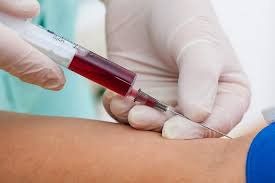
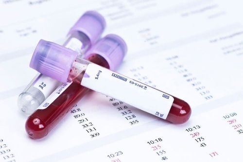
Peripheral blood from the patient serves as the specimen for this test. Healthcare workers will collect 2 ml of peripheral blood, which is then anticoagulated with EDTA.
The patient may feel nothing or only a brief sting when the needle penetrates the skin and vein. The discomfort of blood collection depends on the healthcare worker's skill, the condition of the vein, and the patient’s sensitivity to pain.
4.Technical procedure for the CD3, CD4, CD8 immune cell count test
Put 100μl of anticoagulated peripheral blood into a flow cytometry tube and label it with the patient's name. Add 20μl of each antibody: CD45-PC5, CD3-ECD, CD4-PE, and CD8-FITC, then mix well. The sample is incubated at room temperature for 20 minutes, avoiding light exposure.
Add 2000μl of red blood cell lysis solution and incubate at room temperature for 10 minutes. Immediately after incubation, add 100μl of reference particle suspension, mix well, and analyze using a flow cytometry machine.
Blood samples are analyzed using the pre-installed T-CD3, T-CD4, and T-CD8 count programs on the flow cytometry machine.
5. How long until the results are available?
The CD3, CD4, and CD8 cell count test results are typically available on the same day. Immunology test results are automatically generated by the computer, showing the absolute count and percentages of each T-CD3, T-CD4, and T-CD8 cell type, respectively.
What are normal CD3, CD4, and CD8 levels?
Normal cell counts are as follows:
- T-CD3 cells: 800–2300 cells/μL of blood (55–80%)
- T-CD4 cells: 400–1300 cells/μL of blood (35–55%). A decrease in CD4 cell count may indicate the progression of AIDS in the patient.
- T-CD8 cells: 250–800 cells/μL of blood (20–35%)

6. Potential errors in conducting the CD3, CD4, CD8 cell count test
Errors during sample collection may include inconsistent patient information between the information form and the test tube, clotted blood samples, or missing collection time. Necessary information must be verified in case of inconsistency or incomplete data. If the sample clots, a new specimen must be collected.
Lymphocyte regions not presenting in the program window may result from several factors, including insufficient antibody incubation, improper or incomplete procedure steps, inadequate incubation time, poor pipetting technique, or clogged nozzles. In such cases, the test should be repeated while adhering strictly to procedures. The machine should also be checked, flushed, and the nozzle unclogged following guidelines. If the final result only provides the percentage of T-CD3, T-CD4, and T-CD8 cells without their absolute counts, it may be due to the technician failing to input data on white blood cell count in the specimen, and reference particle concentrations. It is essential to enter this data before reanalyzing the incubated sample tube.
Please dial HOTLINE for more information or register for an appointment HERE. Download MyVinmec app to make appointments faster and to manage your bookings easily.
To arrange an appointment, please call HOTLINE or make your reservation directly HERE. You may also download the MyVinmec app to schedule appointments faster and manage your reservations more conveniently.