Article reviewed by: Pediatric & Neonatology Specialists, Department of Pediatrics & Neonatology, Vinmec Hai Phong International Hospital
Intussusception is a common surgical condition in young children, especially breastfed infants. It occurs when one segment of the intestine slides into an adjacent segment, leading to intestinal obstruction.
1. Causes of Intussusception in Breastfed Infants
The exact cause of acute intussusception in breastfed infants is unclear, but several theories are widely accepted:
🔹 Disproportionate growth between the ileum and cecum:
• At 4 months of age, the ileum and cecum are nearly equal in size.
• Between 4–12 months, the cecum grows faster, increasing the risk of intussusception.
Loose or absent attachment of the cecum and colon to the posterior abdominal wall.
🔹 Underlying conditions such as:
• Meckel’s diverticulum
• Small intestine tumors
• Polyps
• Worm infestations
🔹 Mesenteric lymph node inflammation:
• The Bauhin valve in breastfed infants may protrude into the colon.
• Lymphoid tissue enlargement due to infection can obstruct small bowel motility.
🔹 Viral infections causing mesenteric lymphadenitis:
• Higher incidence of intussusception during respiratory infection seasons.
• Adenovirus, enterovirus, and other viral infections are commonly associated.
🔹 Opposing peristaltic waves in the ileocecal region:
• Ileal peristalsis moves forward, while colonic peristalsis moves backward toward the cecum.
2. Symptoms of Intussusception in Breastfed Infants
✔ Severe, Colicky Abdominal Pain:
• Infant suddenly screams in pain, twists their body, arches their back, and kicks their legs.
• Pain episodes occur suddenly and last 5–15 minutes.
• Pain causes nighttime awakening, daytime irritability, refusal to play or breastfeed.
• Attacks repeat frequently, making the infant progressively weaker and exhausted.
✔ Vomiting:
• Occurs as early as the first pain episode.
• Initially contains undigested food, later progresses to green or yellow vomit.
✔ Bloody, Mucous-Streaked Stool:
• May appear immediately or within 24 hours after onset.
• Stools may be red or brown, containing blood-streaked mucus.
• In some cases, fresh blood droplets may be visible in the diaper.
✔ Bowel Obstruction Symptoms:
• Complete obstruction leads to absent gas and stool passage.
• Symptoms may be mistaken for diarrhea-related complications.
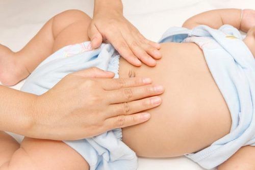
✔ Physical Examination Findings:
• Palpable sausage-shaped mass in the right upper quadrant or above the umbilicus.
• Soft abdomen with no pain between episodes.
• The mass is firm, elongated, and mobile, located along the colon’s path.
Rectal examination may reveal blood-streaked mucus or the leading edge of the intussusception (in low-lying cases).
Empty right iliac fossa due to the cecum’s upward displacement.
✔ Complications After 48 Hours:
• Systemic symptoms:
Early stage: Minimal systemic changes.
Late stage: Lethargy, dehydration, electrolyte imbalance, infection, fever.
3. Diagnosis of Intussusception
✔ Clinical Examination & Imaging Tests:
• Diagnosis is primarily based on clinical signs and imaging, particularly ultrasound.
✔ Plain Abdominal X-ray:
• Limited diagnostic value, but may show:
• • Soft tissue mass in the right upper quadrant or epigastric region.
• • Absence of gas in the right iliac fossa due to cecum displacement.
• • Fluid-air levels indicating bowel obstruction in late cases.
• • Pneumoperitoneum, suggesting bowel necrosis or perforation.
✔ Barium Enema X-ray (Not for Late-Stage Cases ≥ 48 Hours):
Characteristic findings:
• Cup-shaped sign
• Crab-claw sign
• Hook sign
• Target sign
• Cloverleaf sign
✔ Air Enema:
• Shows classic intussusception images, similar to barium enema.
• Safe and widely used in diagnosis.
✔ Ultrasound – Most Reliable Diagnostic Tool:
• Clearly visualizes the intussuscepted segment and its location.
Characteristic Findings:
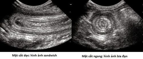
- Nested block image:
Cross-sectional view:
• Round or oval mass, >3 cm in diameter.
• Hyperechoic center surrounded by a hypoechoic outer layer.
Longitudinal view:
• “Sandwich” or “Three-layered” appearance.
• Central bright layer with darker outer layers.
Doppler Ultrasound:
• Assesses intestinal blood flow to determine whether surgery or enema reduction is needed.
• Absence of blood flow suggests bowel ischemia, requiring immediate surgery.
• CT Scan (Rarely Used in Children):
May show small bowel obstruction, with dilated loops above and collapsed loops below the intussusception.
⚠ Parents should seek immediate medical attention if their child shows signs of intussusception.
⚠ Delays can lead to bowel necrosis and life-threatening complications.
For more health, nutrition, and beauty tips, visit Vinmec International General Hospital to safeguard the health of yourself and your loved ones.
To arrange an appointment, please call HOTLINE or make your reservation directly HERE. You may also download the MyVinmec app to schedule appointments faster and manage your reservations more conveniently.